
Frontispiece
Preface
This volume is a corrected shorthand report of the course of lectures adapted to a juvenile audience given by me during the Christmas holidays 1903-4 at the Royal Institution, London. The lantern slides which I used in the lectures have been converted into process blocks. Many of these were photographs specially prepared under my direction for the lectures, and are from specimens in the Natural History Museum. My desire was, as far as possible, to illustrate what I said by photographs taken from actual specimens. Some of these have come out fairly well as process-blocks. For several of the slides and figures I have to thank my friend and colleague Dr. Arthur Smith Woodward, Keeper of the Geological Department of the Museum, to whom I am greatly indebted for kind help in many ways in regard to these lectures. I have also to thank other friends for the loan of lantern-slides and consequent process-blocks, viz., Mr. R. Lydekker, Dr. Bather, Dr. Andrews and Mr. Pyecraft of the British Museum, and Professor Sollas of Oxford. I am also indebted to the Trustees of the British Museum for permission to use several figures of extinct animals taken from the guide-books to the Natural History Museum, published by their order, to Messrs. Macmillan & Co., and to Mr. John Murray.
I trust that this volume will not be regarded as anything more ambitious than an attempt to excite in young people an interest in a most fascinating study, and that it will be understood that it does not profess to give more than a peep at the strange and wonderful history of extinct animals.
	E. RAY LANKESTER.
	1905.
Chapter One: Animals Which Have Lately Become Extinct—The Strata of the Earth’s Crust
Extinct animals are animals which no longer exist in a living state. Of course a vast number of individual animals, and men too, become extinguished, or extinct, in the course of every year, every month and every day.
But the extinct animals of which I wish to speak in these lectures are extinct kinds of animals, kinds of animals which no longer exist on the surface of the globe in a living state, although once they flourished and held their own.
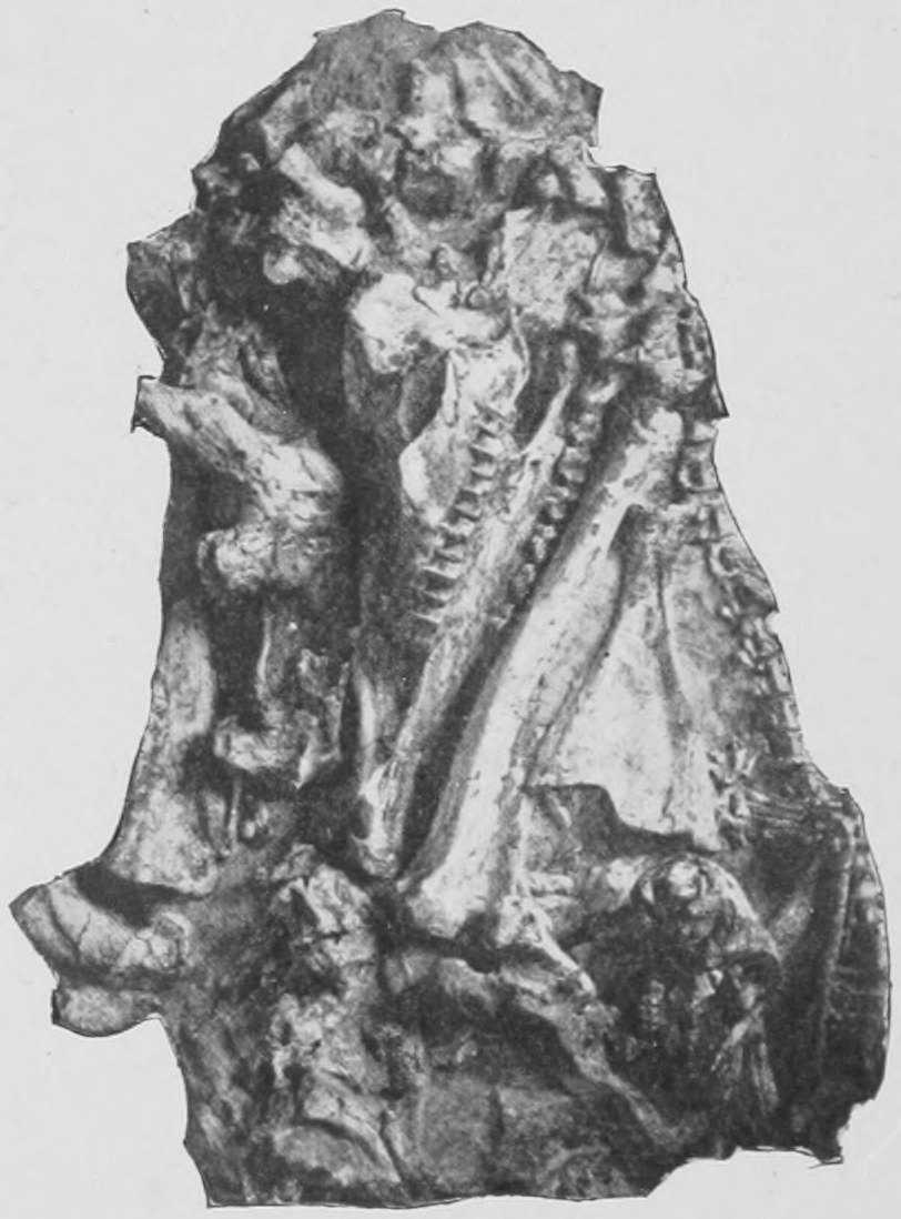
We know of some of them by tradition. The records of men of past ages who have seen some animals, now extinct, and have written about them, and even drawn them, have by human care been passed on to the present day. We know of other extinct animals by finding their bones buried in the ground, some quite near the surface, others deeper in the rocks, far down in the depths of the earth. Such bones may be dug out. There is a sample of such bones found buried in the earth, photographed as our first illustration (Fig. 1). Many of these bones have been so big, so huge, that they have led to the notion of the existence of giants in former days, it not having occurred, apparently, to those who found them, that they were the bones of extinct animals and not of a great race of men.
The indications given by buried remains of a condition of the world which has passed away, as, for instance, in the great buried town of Pompeii, and some of the buried cities of Egypt, excite, when they are dug up, the greatest interest. From the records still preserved to us, we try to find out what was the meaning of the particular objects found, what were the nature and the life of the men to whom they belonged. The same kind of interest belongs to the remains of extinct animals that we dig up, only that many of them are far older than any remains of man ever found. We speak of the remains of an ancient Egyptian city as being some thousands of years old; but the remains of many animals to which I shall have to refer in these lectures have to be estimated, not by thousands of years, but by millions of years; so many years in fact that no numbers with which we are familiar will suffice to bring the facts to the minds of my readers.
Far down in the depths of the earth we find the remains, in a well-preserved condition, of the bones and teeth of such animals; we are able to tell what kind of animals they were, where they lived, what they fed upon, how they moved, and, in fact, their whole general appearance.
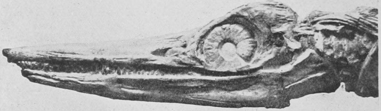
It is urged by some educationists—I myself do not agree with them—that we should present knowledge to young people in a logical order; and that before talking to young or uninstructed people about extinct animals you ought to administer to them a complete course of instruction concerning living animals: that beginners must learn the nature of the structure of living animals, and must study the geography and history of the crust of the ground in which the remains of extinct animals are found, before they can look with any intelligence on extinct animals. That is an opinion which exists. But I do not believe in such a method. The logical method of instruction or study is in my judgment a mistaken one. The whole art of education consists in exciting the desire to know. By showing something wonderful, mysterious, astonishing and marvellous, dug from the earth beneath our feet we may awaken the desire to understand and learn more about that thing. The strangeness of the bones and teeth of extinct animals will lead a boy or girl on to learning about the bones and teeth of living animals in order to make a comparison, and thus to learning more concerning the strange remains dug up. I believe that is usually the case. It certainly was the case with myself. When I was very young, younger than, or as young as any of my readers, I used to be taken by a very kind lady, my governess, to the Natural History Museum of the day, which was then in a remote part of London called Bloomsbury, whence it has been removed to Cromwell Road, Kensington. I was absolutely fascinated as a child with the remains I saw of strange extinct animals. And it is my hope that the boys and girls who read these pages may share some of this interest and fascination, and that they will pass from these lectures to see the actual specimens which are placed on view at the Natural History Museum. These lectures are indeed little more than a sort of invitation to you all to go and see the real things at Cromwell Road, of which I can only show you photographs in this book. I will now show you a portrait of a creature which has always fascinated me with its stony stare. It is the head of an Ichthyosaurus dug out of the rock in the South of England, at Lyme Regis, many years ago (Fig. 2). The eye is peculiarly well preserved. The circle of bony plates, similar to those found in the eyes of birds, give an expression of interest which few fossils can boast of. It was dug out of the rocks by a wonderful lady, Miss Anning, who at the beginning of the last century secured a great number of such remains in the cliffs on the sea-shore. For many years the front part of this specimen was missing, but eventually it was found and dug out of the rocks. I shall have more to say later about creatures of this kind.
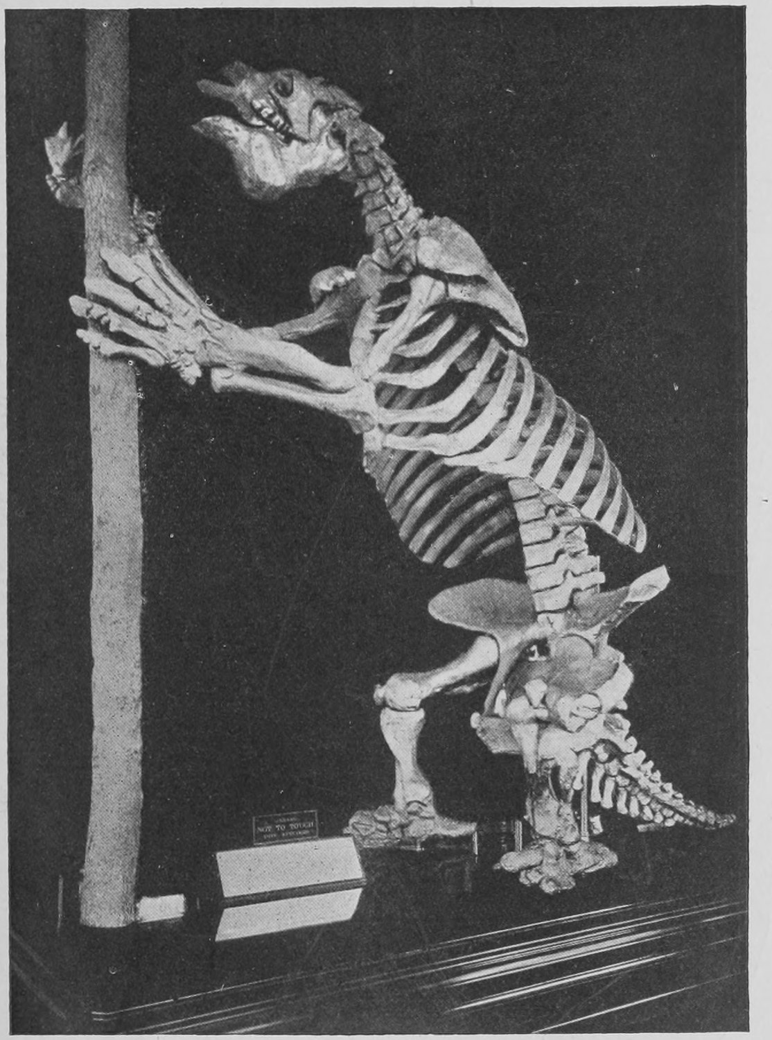
Another creature which fascinated me is shown here as it is exhibited in the East court of the Museum (Fig. 3). It is similar in structure and nature to the sloth. But instead of living on a tree it stood on the ground, and pulled the tree down to it, in order to feed on the young branches. The skeletons of a great many of these huge sloths have been found in the gravel of South America.
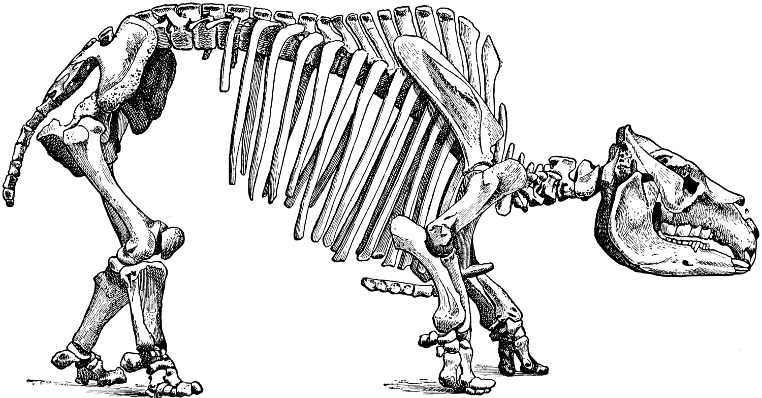
Another strange great creature is revealed to us by this skeleton (Fig. 4), like a huge guinea pig with tremendous chisel-like teeth in front. It also is found in South America. This is the Toxodon.
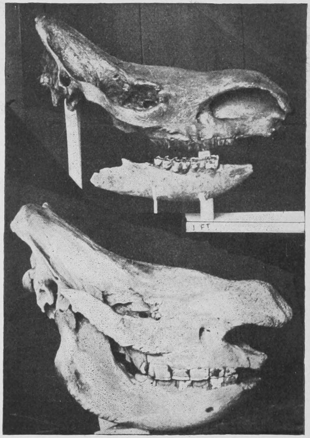
The next picture (Fig. 5) I have here shows the skulls of two rhinoceroses. The lower one is the skull of an African rhinoceros, a living beast known as the square-mouthed or white rhinoceros—called white apparently, not because he is black, but in spite of the fact that he is black. As a matter of fact he sometimes has a number of white patches. But it suffices to know him as the square-mouthed rhinoceros.
The upper specimen is the skull and lower jaw of a rhinoceros, dug up last year in the City of London in Whitefriars, under the office of the well-known newspaper the Daily Chronicle. Digging in the mud and clay there, the workmen came upon this rhinoceros skull. Many such have been found in English river gravels, and we know accordingly that such animals used to exist on the banks of the Thames many thousands of years ago. That specimen also is in the Natural History Museum.
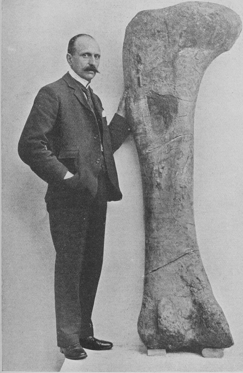
Here (Fig. 6) you have a thigh bone; you can see how enormous it is from the figure of the full-grown man beside it. That is the thigh bone of a huge kind of reptile, bigger than the ordinary elephant, or the biggest African elephant, without counting the reptile's tail. Such remains have been found in England; but the largest have been found in the United States.
These are just a few samples of the remains of extinct animals, and indicate the kind of creatures I want to tell you about. Of course I cannot in these pages refer to all the many thousands of kinds of extinct animals which are known; I can only hope to show you pictures of a few samples of these things, which, however, I hope will suffice to induce you to look further into the matter, to look at the real specimens, and to read more elaborate books, and thus come to feel the same interest and pleasure in examining them that I do myself.
The world upon the surface of which we live has been for millions of years always changing. Nothing is to-day as it was even one hundred years ago. A thousand years brings about enormous changes, quite a different state of things in fact. There are now cities where forests were growing. Animals which existed a thousand years ago have altogether gone. And this history of change has been going on, not merely for a thousand years, but for hundreds and thousands and millions of years. The changes have been incessant, and have been very great.
The difficulty in this study of extinct animals and in the geology connected with it is to think of long enough lapses of time. If you look at that clock you cannot see the hand moving, and yet it is moving. And thus even in a human lifetime you will hardly notice any difference in the rivers and the sea-shore and the cliffs. But if you range over a long enough time, say a thousand years or several thousands of years, and compare the condition which existed a thousand years ago with what exists to-day you will be able to observe great change. The difficulty is to realize this change, for it comes about too slowly for our short lives to give us any real definite experience of it, just as we fail to see the hands of the clock moving when we glance at them for a second. Throughout these lectures I want you always to bear that in mind.
We know of animals even now which are becoming extinct. In this country we have historical records of animals that have become extinct. I will show you one which used to exist in this country.
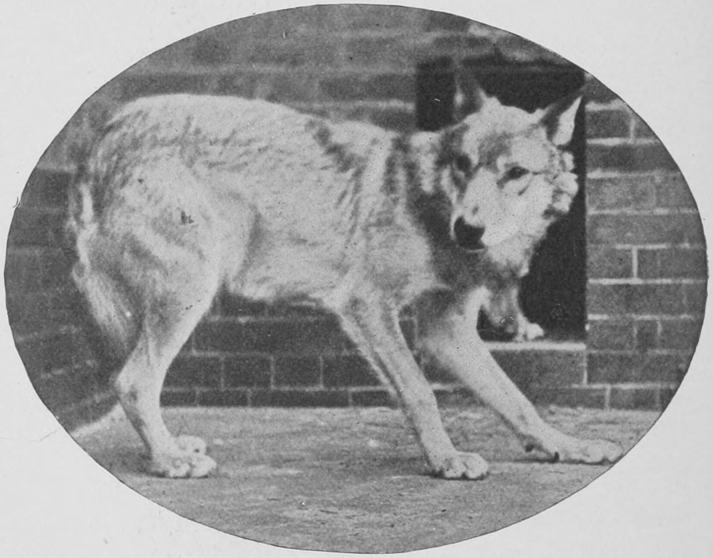
This creature, the grey wolf (Fig. 7), existed in England till the time of Henry VIII., at the end of the fifteenth century, and 150 years later in Scotland and Ireland. But it was entirely exterminated by human beings, on account of its rapacious and dangerous habits. Though it is extinct in England, it still exists in France, Spain, Germany and Russia.
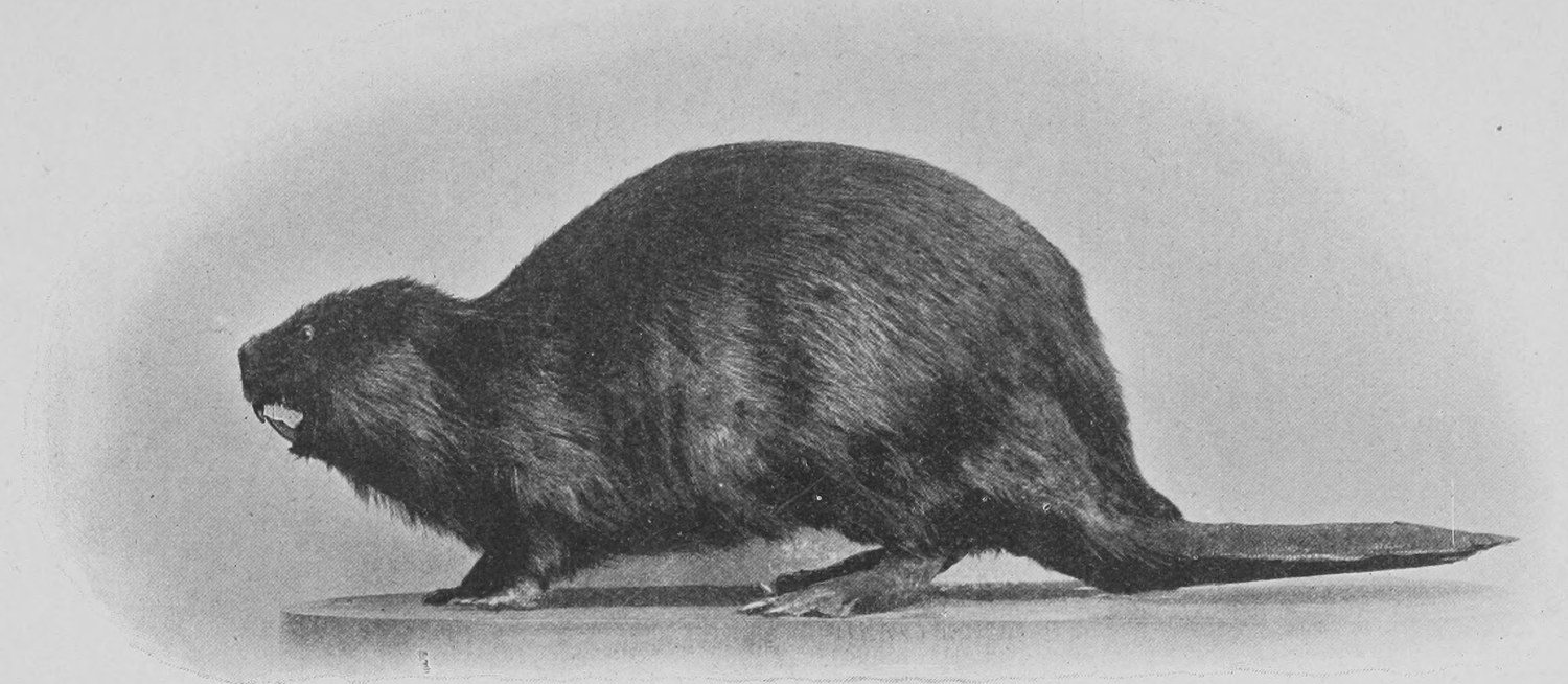
Here is another animal (Fig. 8), the beaver, which used to exist in England, and was found as late as the sixteenth century in Wales. It still exists in France, on the banks of the streams at the mouth of the river Rhone; also in Russia and Scandinavia. In America, in Canada, beavers are still more abundant.
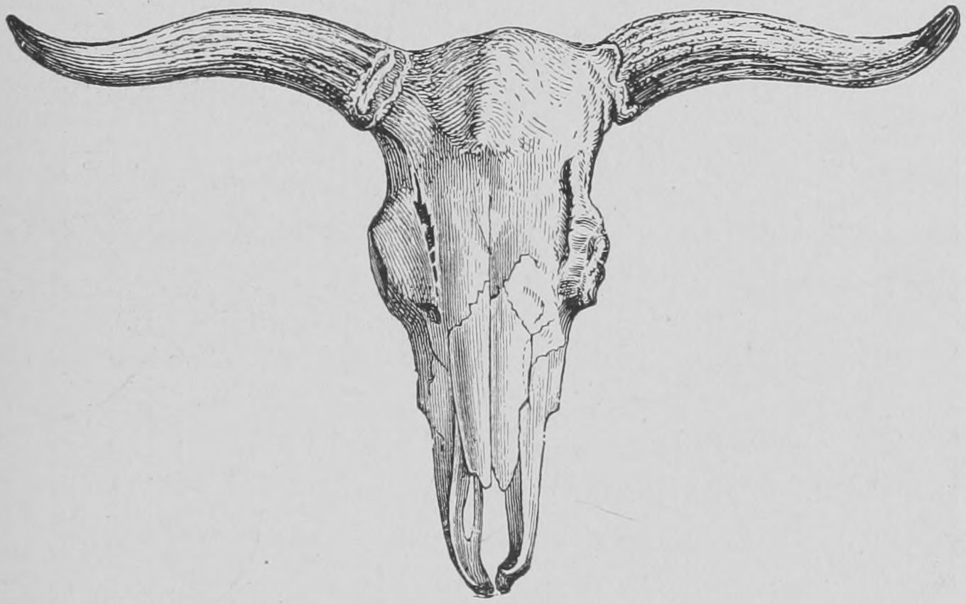
Another creature which, records tell us, existed all over Europe, and which has ceased to exist, is the great bull or Urus of Julius Caesar (Fig. 9). He mentions it as existing wild in different parts of Europe, and says it was nearly as big as an elephant. Well, no such great wild ox now exists in Europe. The last was killed near Warsaw in 1627. All we have now are the breeds derived partly from this, partly from other kinds of bulls, which, are quite changed in their general appearance. Some of the more or less wild cattle in different parts of England, for instance those on Lord Tanqueray's and the Duke of Hamilton's estates, are supposed by some persons to be the remains of this race of wild oxen. But this is probably a mistake. They are really the remains of cattle introduced by the Romans, and have run wild. They are not the Urus of Julius Caesar, which was a good deal bigger than the largest domesticated cattle, even bigger than the white oxen of Umbria.
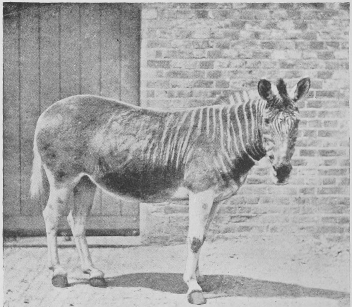
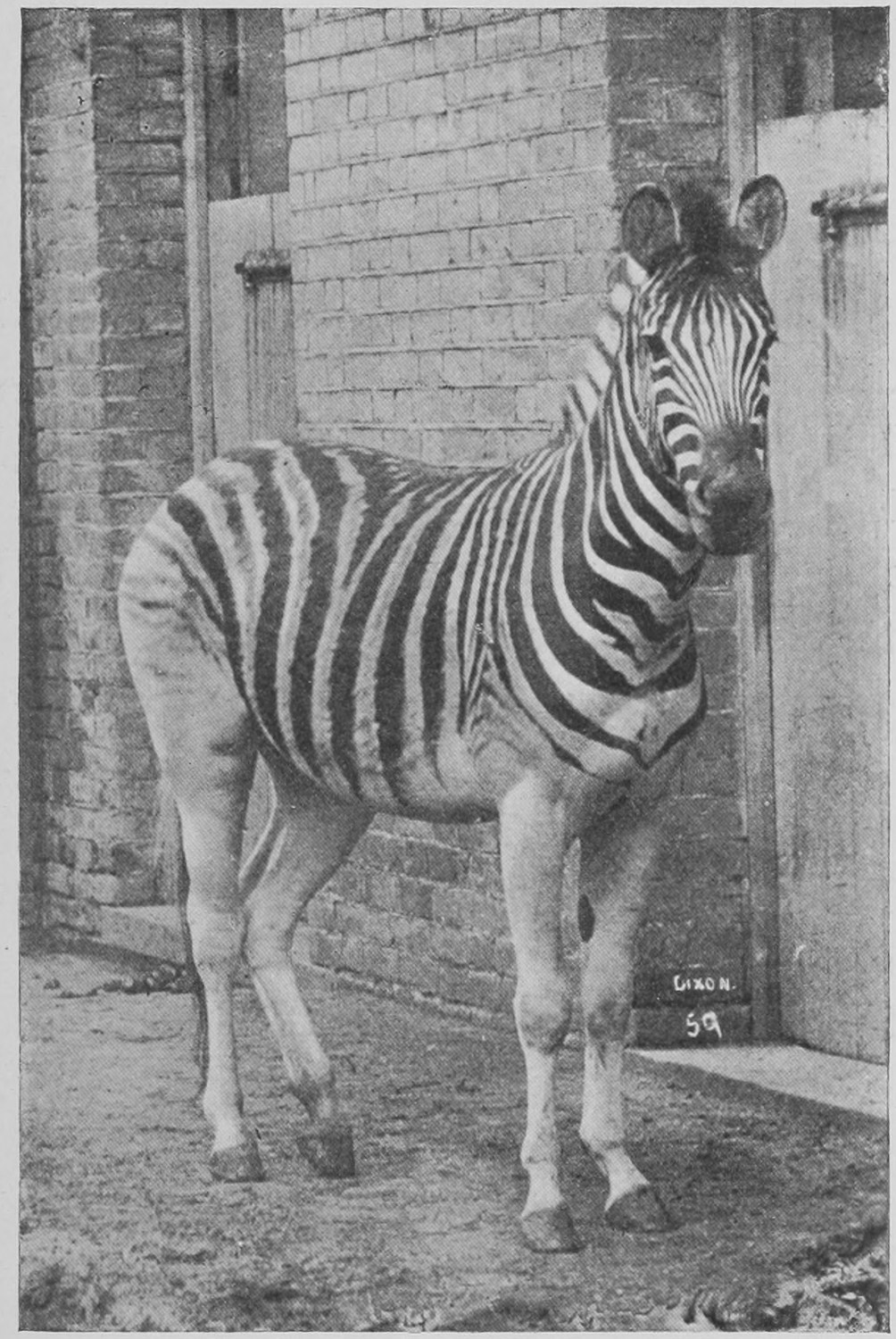
This (Fig. 10) is another animal which has become extinct. But it is not a zebra, as no doubt some of you thought it must be. This is the quagga, which differs from the zebra in being striped in front only. The quagga lived in South Africa, and was quite common there until forty years ago. This photograph was taken from a specimen which lived in the Zoological Gardens some twenty-five years ago. Its stuffed skin is preserved in the Natural History Museum. This creature has now entirely ceased to exist, owing to the fact that the country over which it ranged has been taken up and cultivated by white men. There are no more living quaggas anywhere. This animal has become extinguished in our own lifetime. Zebras (Fig. 11), however, are still common enough in Africa, with their beautiful stripings on the head, and on the fore as well as on the hind regions of the body and legs.
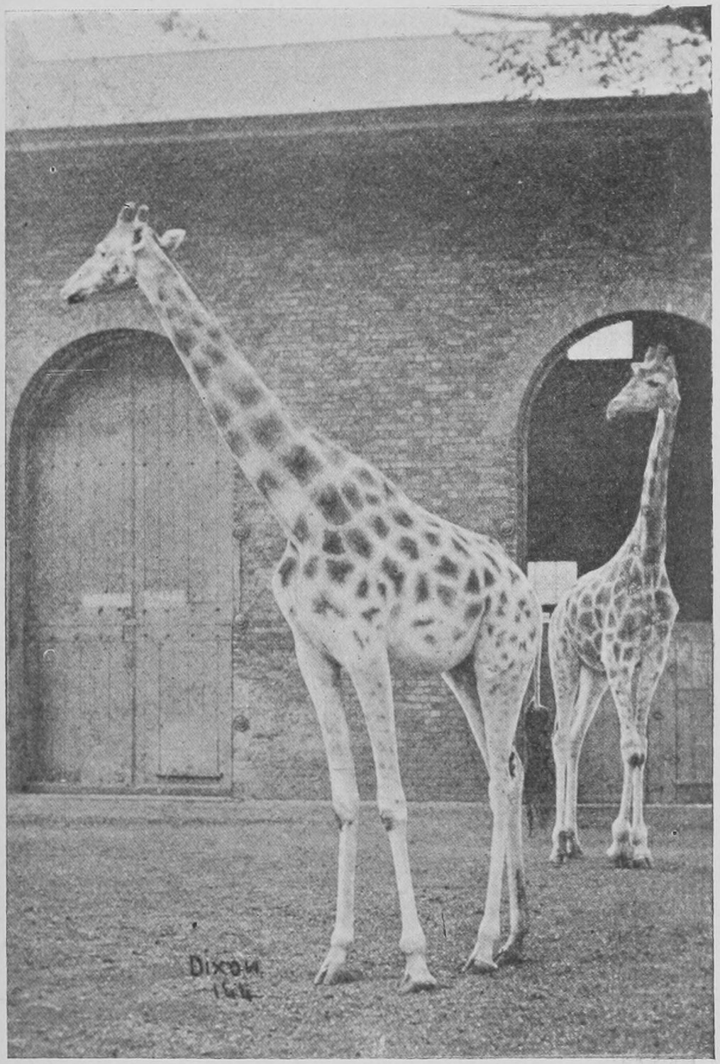
Here is an animal which, it is feared, is becoming extinct—the giraffe (Fig. 12). In South Africa it has become extinct already. But sportsmen now seek it in Equatorial Africa. It is still existing in great numbers in that region, and we hope now will be properly protected by Government. Two new and well-mounted specimens have recently been put in the Natural History Museum. The neck of the giraffe is often represented as growing up from the body with a graceful curve, as is seen in the neck of the swan. But the true position of the neck is as you see here (Fig. 12). The specimens in the Natural History Museum shows this properly.
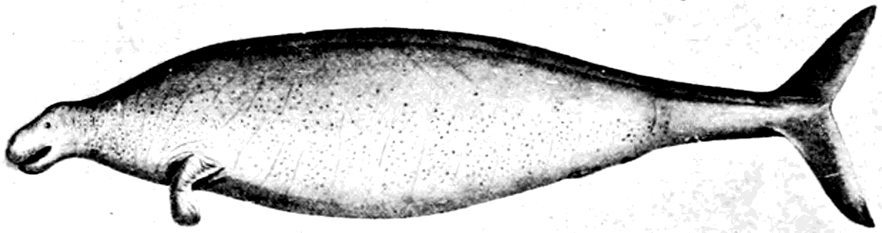
This is a picture (Fig. 13) of a curious creature, an animal known as the sea-cow, found in the Aleutian Islands, between North America and Asia. It was discovered by the traveller-naturalist Steller in the eighteenth century. It was no sooner found than sailors went to the islands where it existed, knocked it on the head and ate it, and in about ten years it ceased to exist. This picture is from Steller's drawing. It is an enormous creature, some twenty feet long, and in shape something like a seal. But it is not in reality a seal or a whale, but belongs to a peculiar group of vegetable-feeding marine animals, the Sirenians. It has a small head, flipper-like fins, no hind limbs, and a fish-like tail.
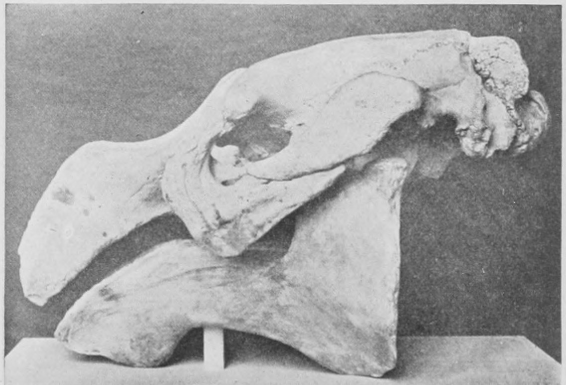
The skull of the same animal is shown in Fig. 14. It has no teeth, but instead bony plates.
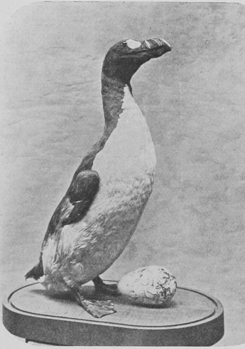
This is the picture of a celebrated animal (Fig. 15)—for you must understand that birds are animals. You will have been handed a list of the groups of animals (see the end of this chapter). I shall not have space to explain it at any length, but it gives the division of animals into groups and their relation one to another. It shows how they are classified, so that I need not refer to the classification again.
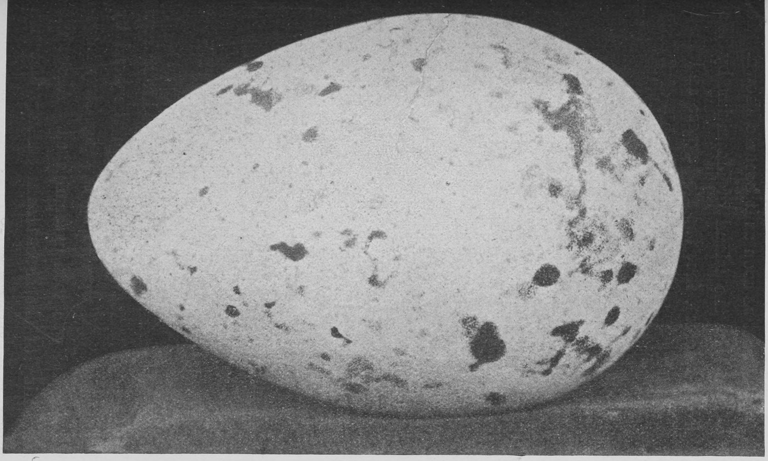
This picture (Fig. 15) is the portrait of an interesting bird, the Great Auk. It is only about 2½ feet high. It is like the penguin in appearance, but it is really related to the puffin and albatross. Fig. 15a shews the egg, which from time to time in the newspapers, we read of as being sold to enthusiastic egg-collectors for as much as £300. Nearly a hundred specimens of the egg of this bird are known, for it only became absolutely extinct some sixty years ago. It used to be found on the rocky islands off the North of Scotland, Shetland, Iceland and Greenland. But it has now absolutely ceased to exist. It is very difficult to say why it died out, for it had not been hunted down. Since it has become extinct we have been able to get to know about it by finding its skeleton buried in sand and guano in certain places on the coast of Newfoundland.
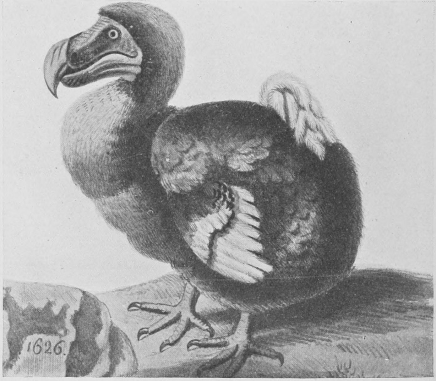
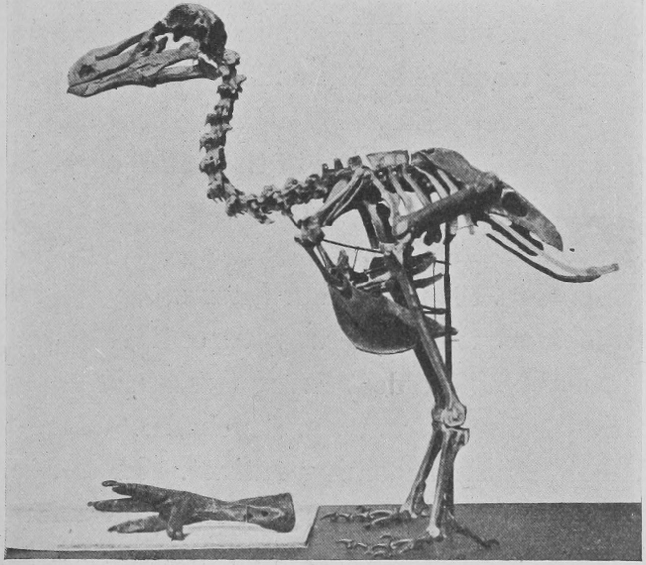
Here (Fig. 16) is another creature, the dodo, a bird which, like Steller's sea-cow, became extinct almost as soon as it became known. It was found in the island of Mauritius by the earlier explorers, first the Portuguese and then the Dutch. The bird was incapable of flying, as it was too fat for its little wings to lift it from the ground. It was knocked on the head by the sailors and worried by the pigs they introduced, and was soon exterminated. About the beginning of the seventeenth century, between 1610 and 1620, specimens were brought alive to Europe and were exhibited as a show. We once possessed at Oxford a stuffed specimen, secured by that ingenious and worthy gentleman Mr. Elias Ashmole, who gave his collections to the university 250 years ago. But as it became mouldy and eaten by insects, it was ordered, a hundred years ago, by the Vice-Chancellor and Proctors of the University of Oxford, that the specimen of the dodo should be destroyed. They do not like mouldy things at Oxford. But the curator cut off the head and one foot, and kept them. This head and foot, together with another foot in London, and a skull in Copenhagen, are about all we have left of dodos seen in the living state by Europeans. But since the dodo became extinct, by digging in the mud of a lake in Mauritius skeletons and bones of it have been found (Fig. 17).
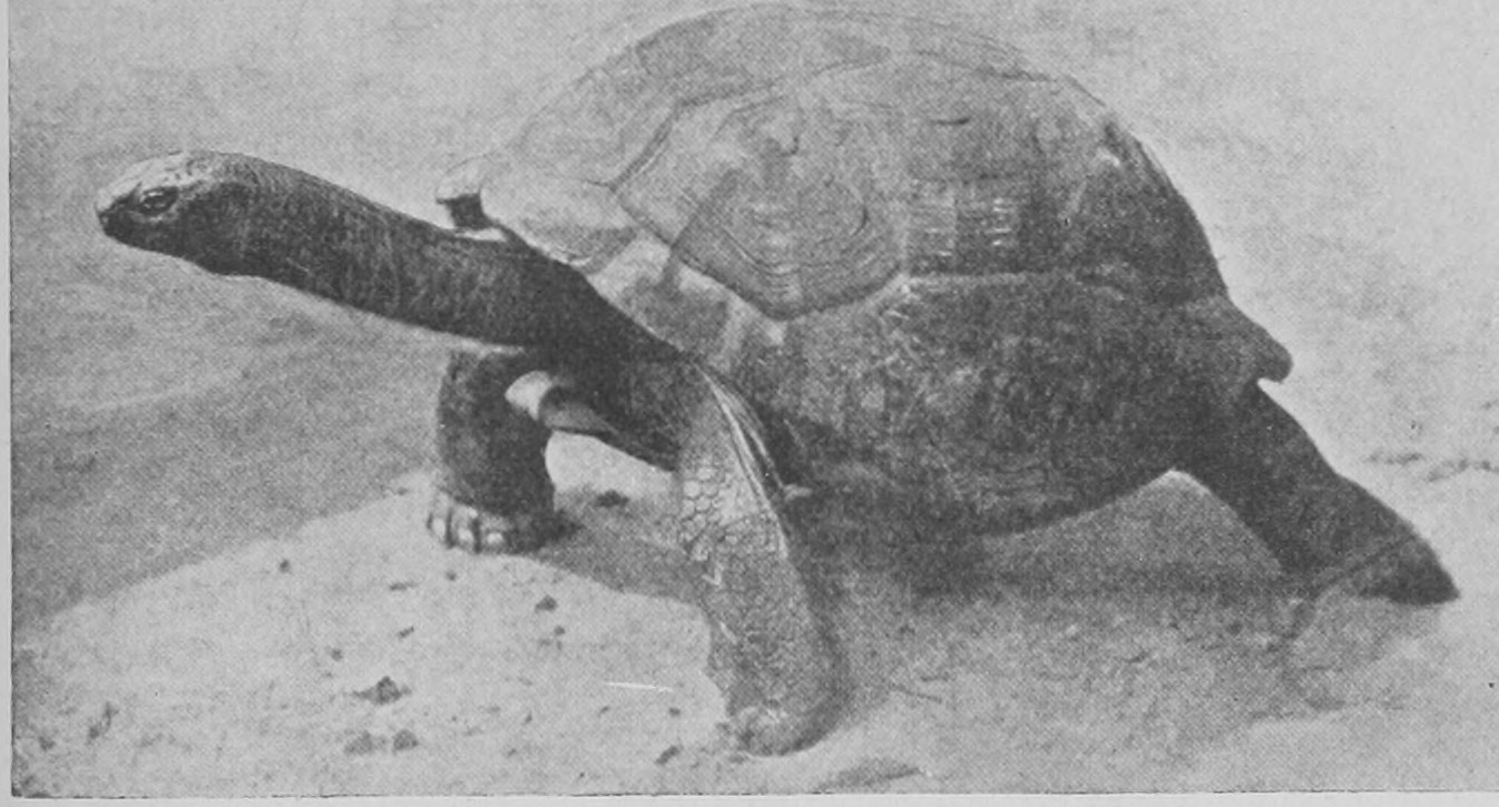
This (Fig. 18) is another interesting creature, whose kind is on the way to extinction. It is probably the oldest living terrestrial animal. It was brought from the Seychelles, where its kind is rapidly becoming extinct. In different oceanic islands such tortoises have been found of large size. This specimen was brought in 1764 to the island of Mauritius, and is still alive there. Thus it has been 140 years in captivity in the Court House Garden, in the Mauritius; and how old it was when brought there it is impossible to say.
A question of great interest is—"What makes animals become extinct?" It is obvious in many cases that another animal, Man, interferes. He either kills and eats animals, or takes their food from them, or occupies their ground, or cuts down the forests in which they live, and so on. But before man appeared on the scene there were changes going on, and different kinds of animals succeeded one another. We know this by finding the remains of different animals at different depths in the crust of the earth, in the different strata which have succeeded one another. The cause of these changes, the cause of the extinction of animals, is a very elaborate and difficult question, and one which I do not propose to deal with at any length. It is connected, of course, with the whole doctrine of the origin of the different kinds of animals. We all recognize now that there has been a gradual development of the different forms of animals by natural birth, from ancestral forms more or less like themselves. But the more remote we get from the present day, in the line of descent, the less like are the ancestors to the present form. The original parental forms have given rise to very different branches of descent. The descendants of one ancestral form have branched out in different directions: just in the same way as some person named Smith at the time of the Conqueror has given rise to all sorts of Smiths. Some of them perhaps are still actually metal-workers, others have become Ministers of State and Right Honourable judges; others have great possessions; but they can all be traced back to the one original Smith. So many living animals of various appearance and form can be traced back to one ancient ancestral form, and these again to other more primitive ancestral forms.
The reason why the ancestral forms died out is really connected with the general change in the surface of the earth. New forms have gradually taken the place of the old forms—for no piece of land remains the same for many years. A thousand years, as I have said, in this matter is merely nothing, but even in a thousand years we get great changes in the surface of the land. Land may rise far above the sea, and what was an island become part of a continent. And what was part of a continent may partly sink, and become an island—that is, the connexion between it and the continent may become covered with water; and then the conditions of life for the animals are very much changed. Such currents as the Gulf Stream are affected by this alteration in land and water. Were certain changes to take place, the warm water of the Gulf Stream would no longer warm certain land; the climate would become colder than the animals have been accustomed to. The animals that could not stand the cold would die out, whilst those that could stand the cold would flourish.1 All I would say is that changes in the disposition of land and water have been a great cause in changing the forms of animals and in bringing about the extinction of one set and the flourishing of another set. That this rising and sinking of the surface of the land really takes place I will try now to give you evidence.
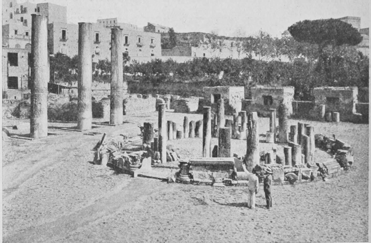
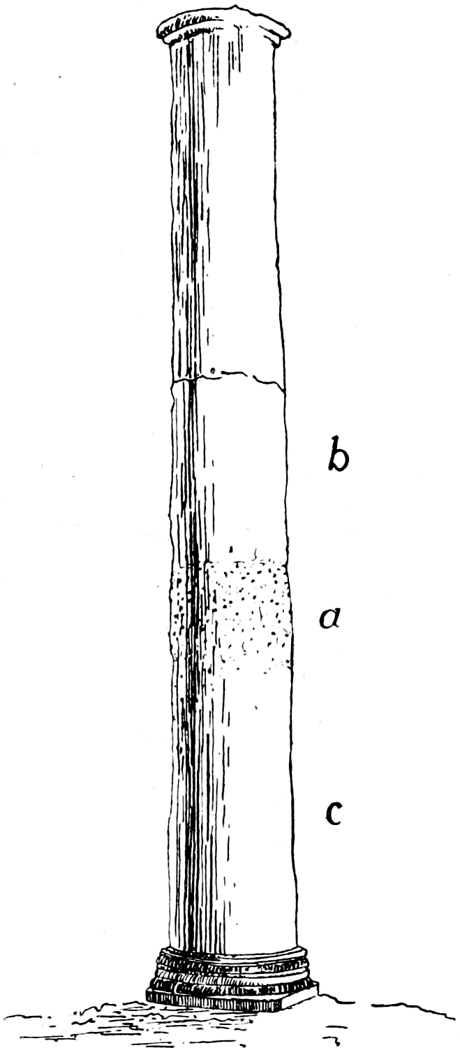
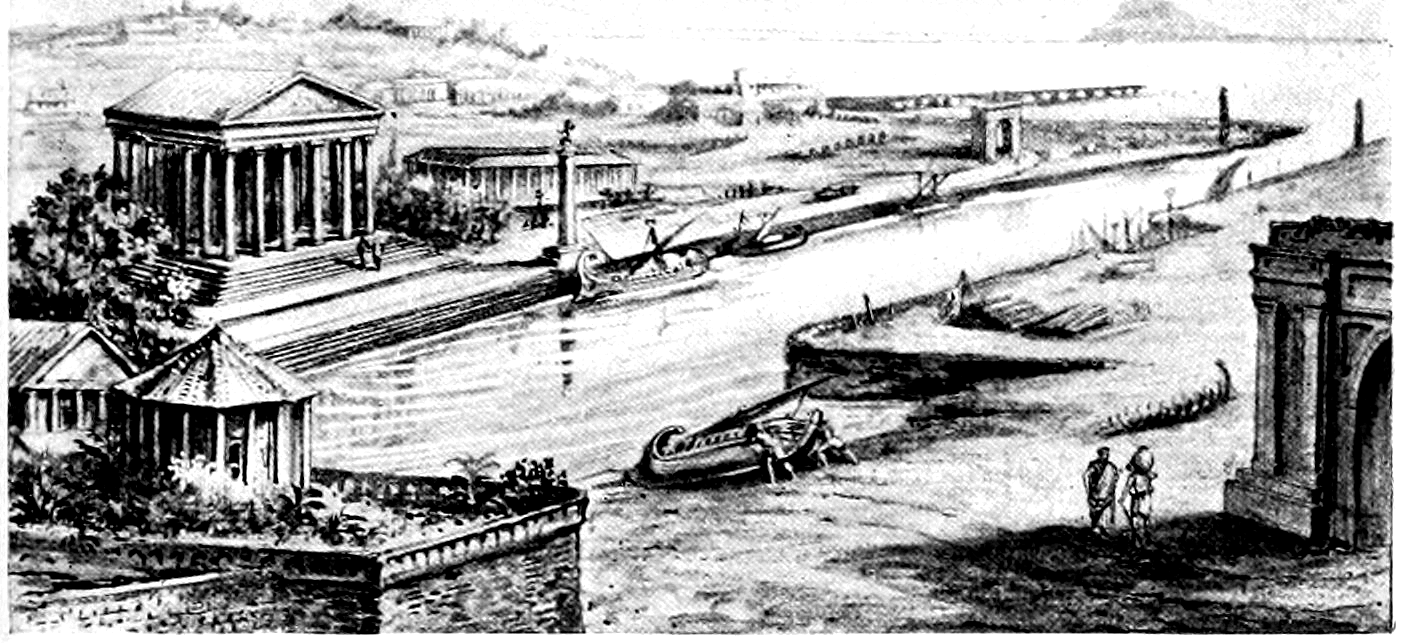
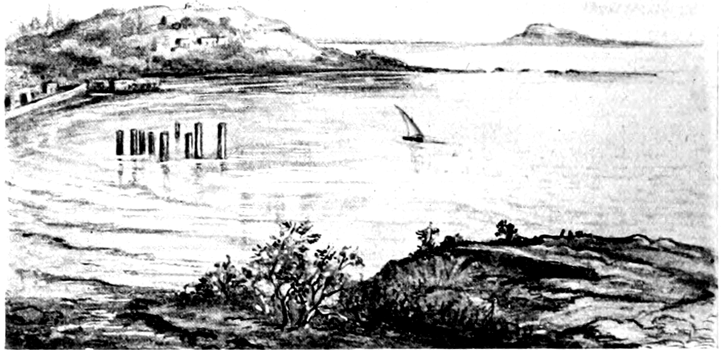
Here (Fig. 19) is the photograph of the Temple at Puzzuoli, near Naples, on the shore of the Gulf of Naples. This has been celebrated for something like eighty years, ever since Mr. Babbage carefully examined and described it, and thus caused it to be largely visited by geologists. In common with most geologists, I have had the pleasure of visiting it. The three standing columns have marks of discoloration up to a certain height. The lower part, as shown in the diagram (Fig. 20), is full of little holes in which tiny sea creatures have burrowed holes in which there are small shells. This is so defined that it is certain these columns have stood in sea water up to that line. The evidence of that is quite complete. These columns formed part of a Temple or public palace in the great Roman town of Puteoli, which had in front of it a Roman road along by the sea-shore. Between the temple and the sea was the road. Now in Roman times that temple stood complete and very much in the same position relatively to the sea-level that it does to-day, but rather higher up. Mr. Günther, of Oxford, examining the shore-line carefully, has found covered over by the sea the remains of the Roman road, and the remains of great blocks to which ships were moored when they brought their wares to the town of Puteoli. I have here made a drawing of the town and the great public palace as it must have appeared in Roman times (Fig. 21). In the distance is the island of Nicida; in the foreground we have the palace and the town, the quay and harbour. Things existed thus in the days of the Roman empire, in the third century of our era. Then earthquakes occurred, the columns were broken, the city sank beneath the sea. We have no written history of this town. But it is known that in the Middle Ages, in the eighth or ninth century, the whole of the coast of this part of Italy had sunk many feet, and the columns were broken and standing in the sea. This is the appearance then presented by Puteoli (Fig. 22). The coast had sunk; the remains of the road were covered by sea, and also the remains of the columns up to the height marked a on the diagram (Fig. 20). The whole land must have sunk as much as forty feet, since the temple or palace stood on high ground originally. Then it was that, while they were under water in the ninth century, the columns were bored into by sea-shells. Now some more centuries have elapsed; the ground has risen again until we have the condition shown in the photograph (Fig. 23), which gives a general view of the same region as that drawn by the use of the imagination in Figs. 21 and 22. The land rose again from the water. But the sea left its mark on the columns, showing exactly how deeply they were merged in the intervening centuries.
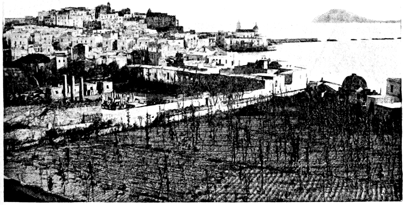
This is considered one of the clearest and most direct proofs of the changes which take place in the level of the ground. The change need not be a continuous or a rapid one. It took some two or three centuries for that temple to sink into the water, and a few more centuries for it to come out again.
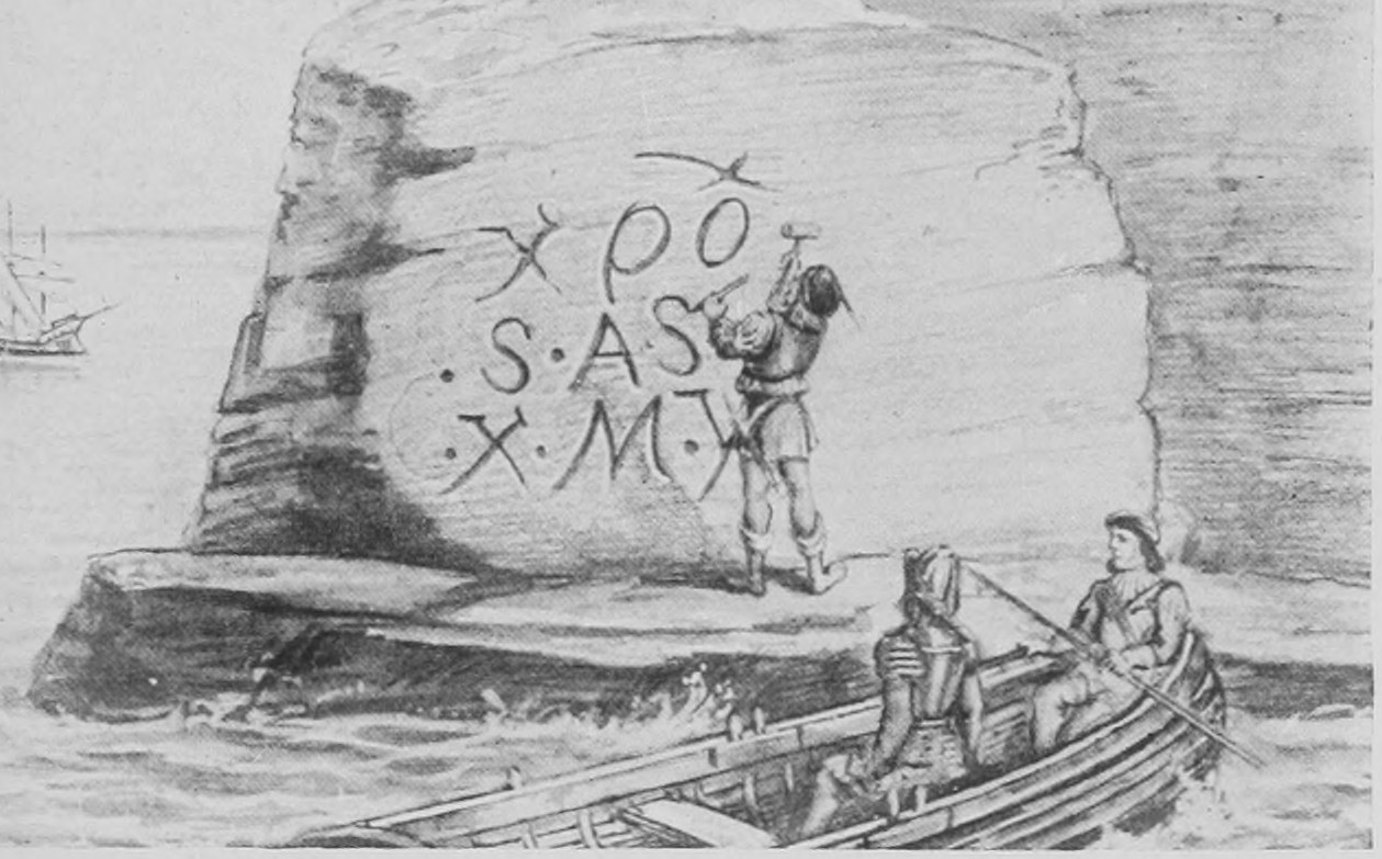
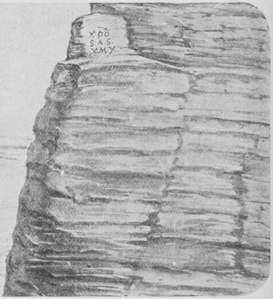
Such movement is always going on. It does not occur very obviously on our own coast. It can be seen to some extent on the Devonshire coast at Plymouth. You get evidence of it in what are called raised beaches above the level of the ocean. In Norway this kind of thing is very obvious. In South America it is going on, and has been going on at an enormous rate for the last thousand years. Probably a great part of the height of the Andes has been acquired within the last few thousand years by rapid rising. When the original sailors landed on the coast of Chili in the sixteenth century or thereabouts they are said in one spot to have chiselled on the rocks an inscription. Here you see an imaginary sketch of them doing so (Fig. 24). It is said, but I cannot find any accurate record of it, that such inscriptions have been discovered now, raised high up on the cliff (see Fig. 25). We know that many kinds of sea-shells are found 200 and 300 feet up the cliffs in this part of the world. According to the observations that have been made, the original inscription which we see the sailors cutting in Fig. 24 would, after 300 years, be found high and dry some 150 feet up the face of the cliffs. On the coast of South America there is good reason for believing that a movement upwards goes on at the rate of half a foot to a foot a year. If such a rising continued for a thousand years we should find that the original shore-line had risen 500 feet above the sea-level.
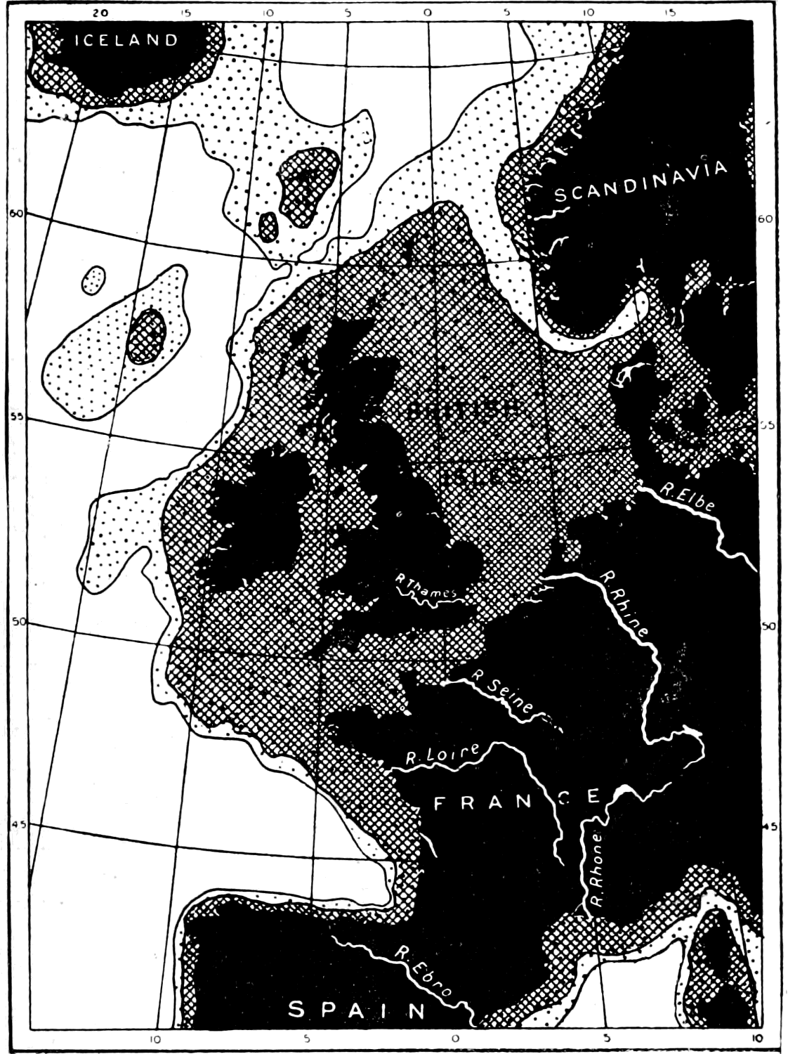
What, then, is the general result of such movement? I will show you what would be the result of elevating the shore of England (the whole of this part of the world) 600 feet. From this map (Fig. 26) you will see that if the floor of the ocean were raised 600 feet, the cross-shaded area would become dry land, and we should be brought by land into contact with the neighbouring continent and islands. And if the land were raised 3,000 feet we should have a still greater extension of dry land. Even the smaller change would make England part of the Continent of Europe.
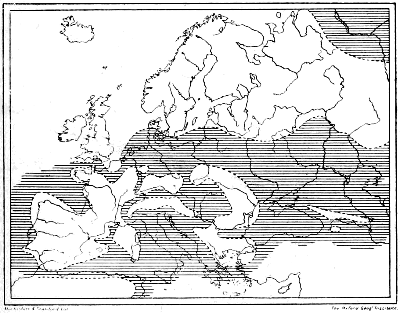
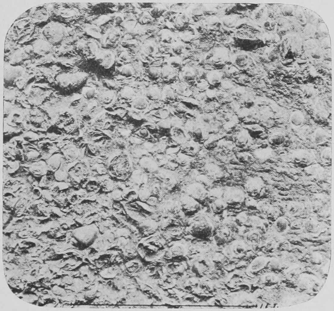
The study of extinct animals found in the various strata of the earth enables one to arrive at a notion of the distribution of land and water in past time. Here is an arrangement of land and water which we are able to conclude must have existed in Europe in what is called the Middle Tertiary period (Fig. 27). All this darker part is the sea, and the pale part land: in fact, the distribution is quite different from what it is at the present time. The whole surface of the earth has been shifting and changing all through time. During the millions and millions of years of past ages, different seas have arisen, different continents, different dry land and different animals,—changed by the various influences of the land and climate. And all this movement is accomplished by the slow cracking and "curling" of the earth's crust, by the continual washing of the surface of the land by rain and rivers, by the eating away of the edge of the land by the waves of the sea. This "eating away" of the land by the sea—quite apart from any sinking of the land-level—has caused and is yearly causing great loss of land on the east coast of England, especially in Suffolk, where the great city of Dunwich has been swallowed up by the sea. In other parts the sea throws up sand and adds miles of new land to the coast. The immense quantity of stuff which is carried off the surface by rains and rivers is difficult at first to imagine. Taking the river Thames at Kingston, it is found that something like 500,000 tons of solid salts of lime in solution is carried every year past that spot. Now a cubic block of limestone measuring a yard in each dimension weighs about two tons. Accordingly, 250 thousand solid cubic yards of rock are carried past Kingston every year by this little river! Enough to build a new St. Paul's Cathedral every year! Think, then, what must be the enormous quantities of solid matter dissolved and carried away by such rivers as the Mississippi and the Amazon. And remember that in addition to this dissolved limestone there is almost as large a bulk of fine sand and mud carried along by most rivers! What becomes of it? It is deposited in layers, and forms what we call stratified rock. You see it, some of it, on the seashore when the tide goes back, in the form of layers of sand, but most of it is deposited far out in the deep bottom of the sea—the lime being taken out of solution by shell-making plants and animals. But where the land is rising, the sand or ground which is exposed when the tide goes back, would after a few years have been raised away from the sea and become hard rock. Layer after layer is imposed and raised from the sea bottom. Without going into detail we may accept as a fact that this formation of layers by stuff brought down from the land by rivers and washed from the coast-line by the sea waves gives rise to what are called "stratified deposits." I will now show you some pictures of this stratification. Here (Fig. 28) are shells embedded in the Bognor rocks deposited some thousand million years ago; there are many sorts of shells, whelk-like shells and volute shells embedded here.
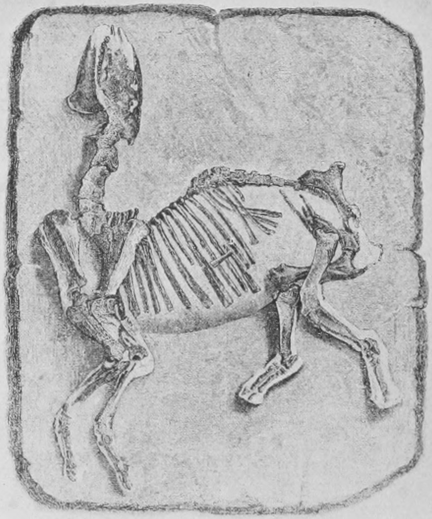
Whole skeletons of animals are sometimes found in the stratified deposits. This one (Fig. 29) is from stratified rock which forms the hills round Paris, the calcareous rocks of Montmartre.
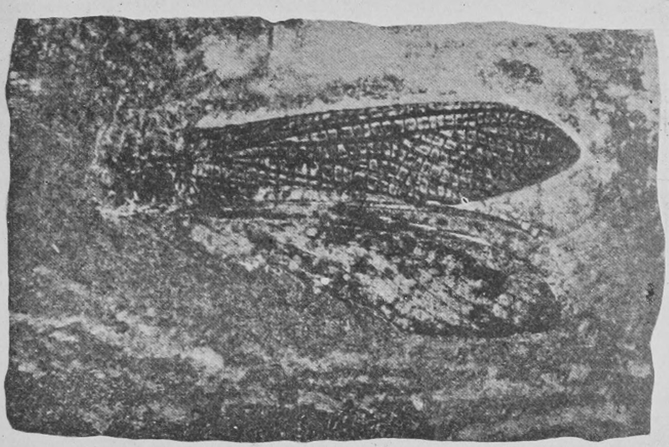
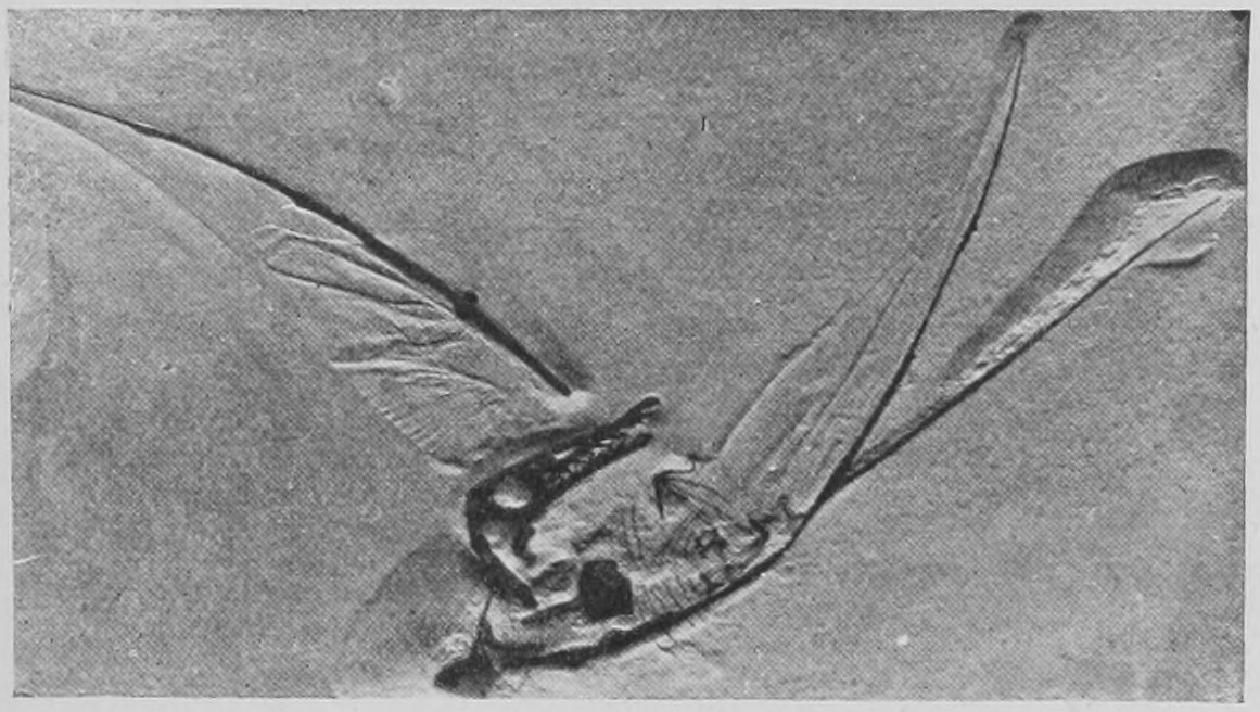
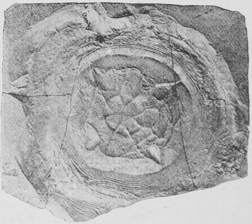
Next let us see what fine mud will do in preserving the impression of delicate structures, such as the wings of insects. Here are the wings of a dragon fly (Fig. 30), preserved in very ancient stratified rock, the Carboniferous. Here (Fig. 31) are the wings of the Pterodactyle preserved in fine sandy limestone of Oolitic age. Here (Fig. 32) we have a jelly-fish preserved; you see its seal stamped as it were on the sand. It is many millions of years old, from Oolitic rock.
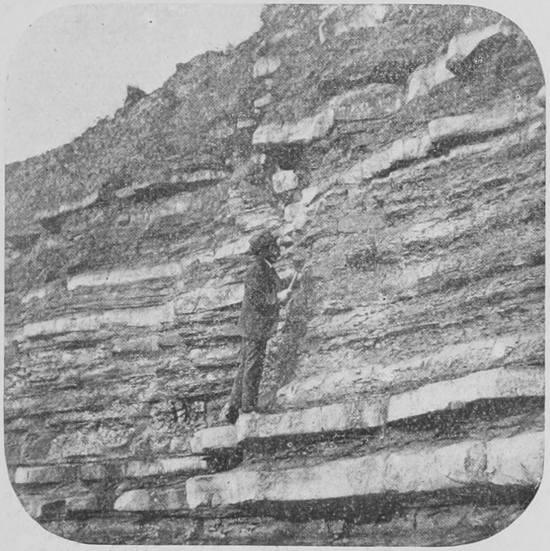
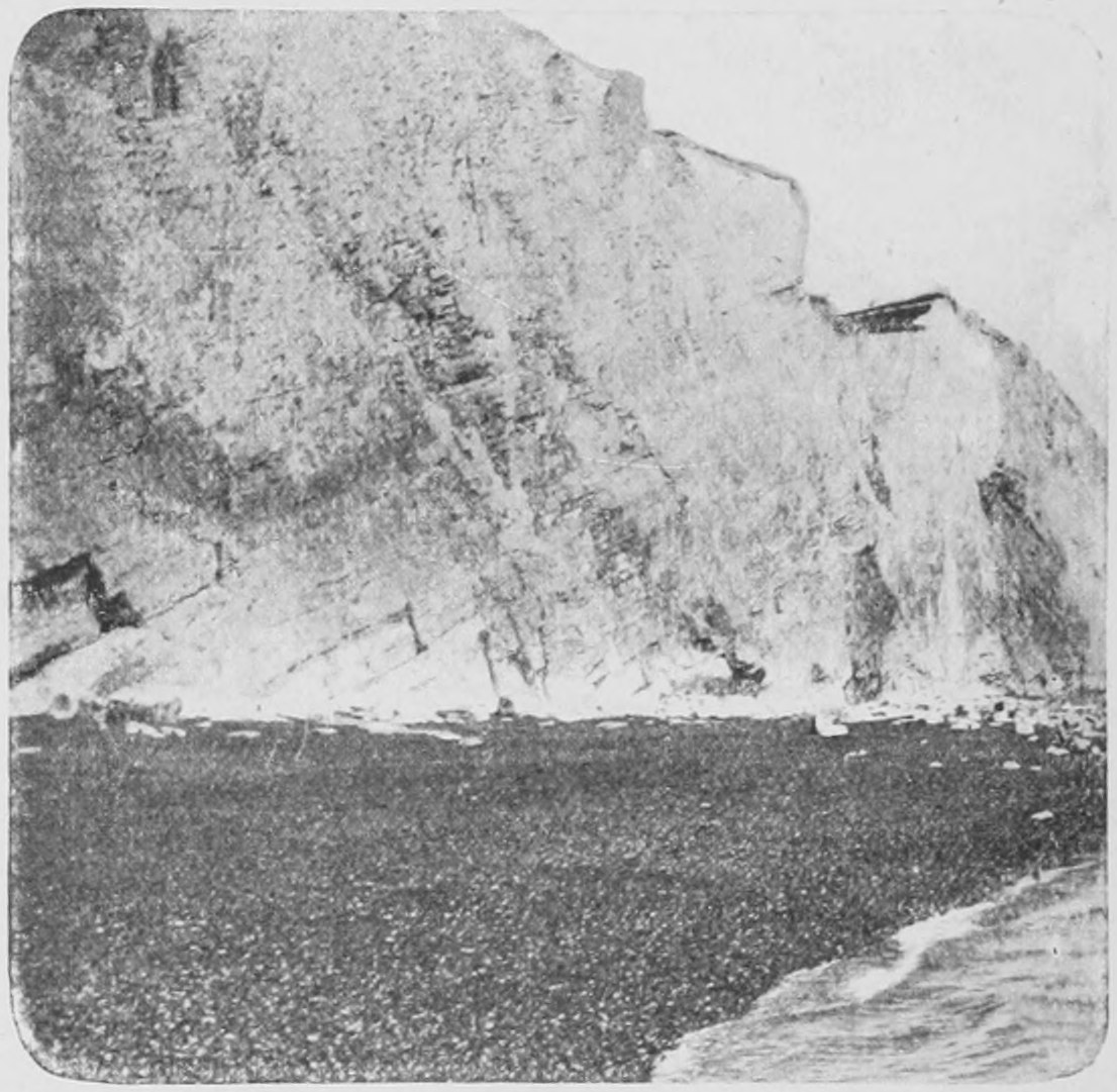
Now let us look at the layers or the stratification of rocks. This picture (Fig. 33) shows part of the cliff at Lyme Regis, where the Ichthyosaurus-head, which I showed you just now, was found. We see the layers of harder and softer material lying one over the other. The next figure (Fig. 34) shows how the layers of the surface of the earth may be bent. Without digging far into the earth you may reach a deep layer of stratification or "stratum" brought near the surface by the general tilting.
This (Fig. 34) is part of the chalk cliff at Seaford, showing the strata tilted, so that the deeper layers come to the surface.
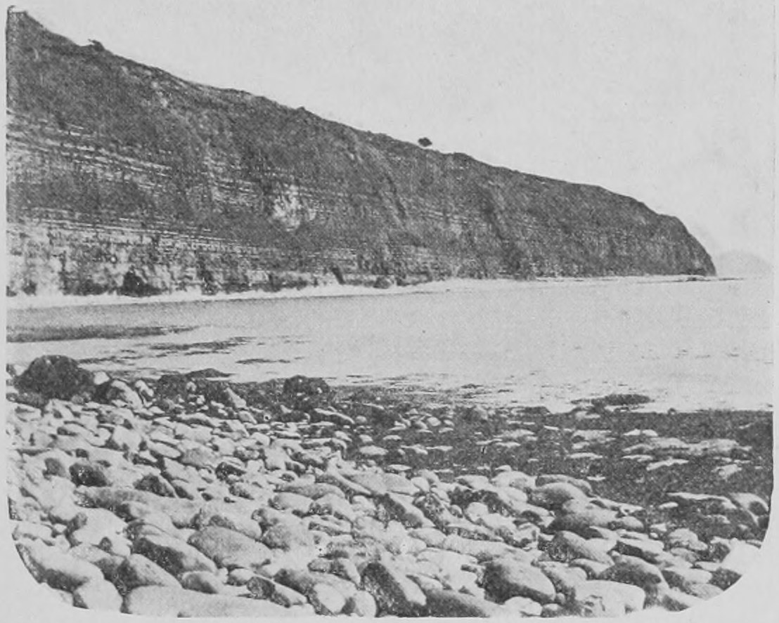
Here (Fig. 35) is part of the shore of Lyme Regis, showing the strata exposed by the action of the sea. A long series of superimposed layers one on top of the other is seen. They are slightly tilted, so that the deeper strata come to the surface near the observer.
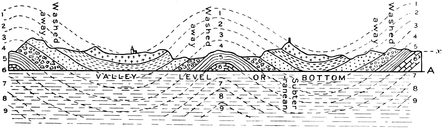
The tilting of the strata of the earth's crust is the rule and not the exception. It is rare for the strata to lie in a strictly horizontal position. The crust of the earth is continually being slowly pushed up or down, and as it were "crumpled" or thrown into wave-like folds. The cause of this crumpling is to be found in the shrinking of the earth and the movements of subterranean steam—causing earthquakes and other earth movements. The "crust" of the earth is a mere skin. If we bored twenty miles into it we should come to immensely hot molten material, and on this the crust is supported. It cannot be said to "rest" on the deeper matter, for it is always, though very, very slowly, shifting and crumpling. Consequently, according to the height and depth of the folds of the crust, we find that deeper, even very deep-lying strata may be brought to the surface, and as the upraised folds get worn away by sea and rain and rivers, the deepest layers may be exposed on the surface. Thus it is that we are able to examine the oldest rocks and to search for the remains of the immensely-ancient creatures which they contain. The diagram (Fig. 36) will help to make it clear how the pushing and crushing of the earth's crust into wave-like folds such as you may see when a tablecloth or carpet is not spread flat, results in bringing the deep-lying strata to the surface, so that we can walk along a cutting or cliff and come to deeper and older rocks as we walk along.
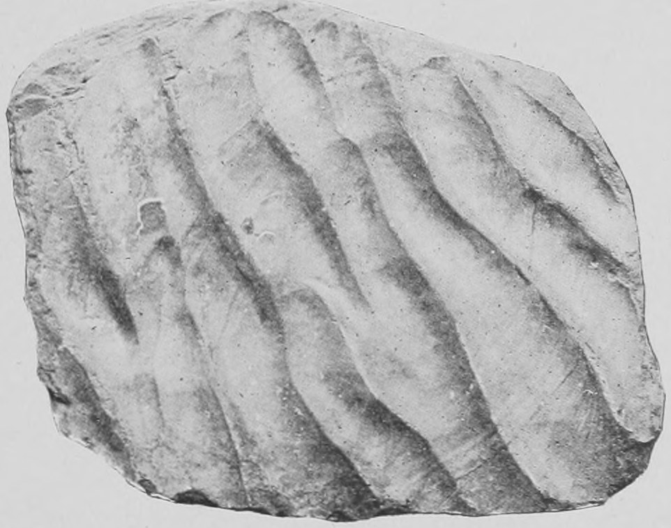
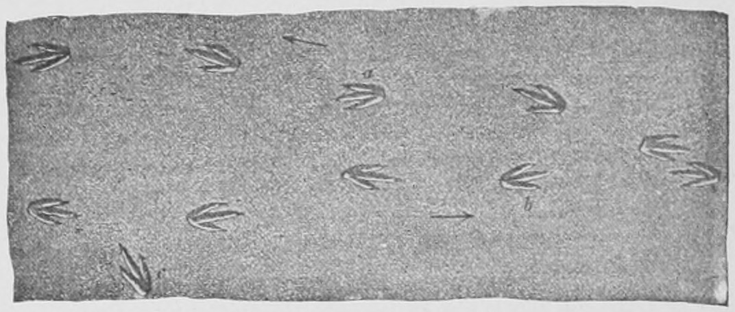
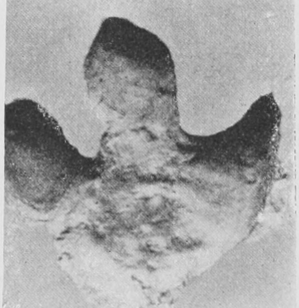
Here (Fig. 37) is a specimen which shows ripple-marks still preserved as we see them nowadays on the shore at low tide. The marks of raindrops are also often preserved on such slabs of rock, which once were soft wet sand. On such surfaces we often find footprints, the footprints of birds and of reptiles. In some cases we do not know the animal itself (Fig. 38), but we see its footprints in the ancient rocks now far removed from the sea and covered over by thousands of feet of later rocks. Here (Fig. 39) are the footprints of a great creature from the Isle of Wight which has left its impress in the sandstone.
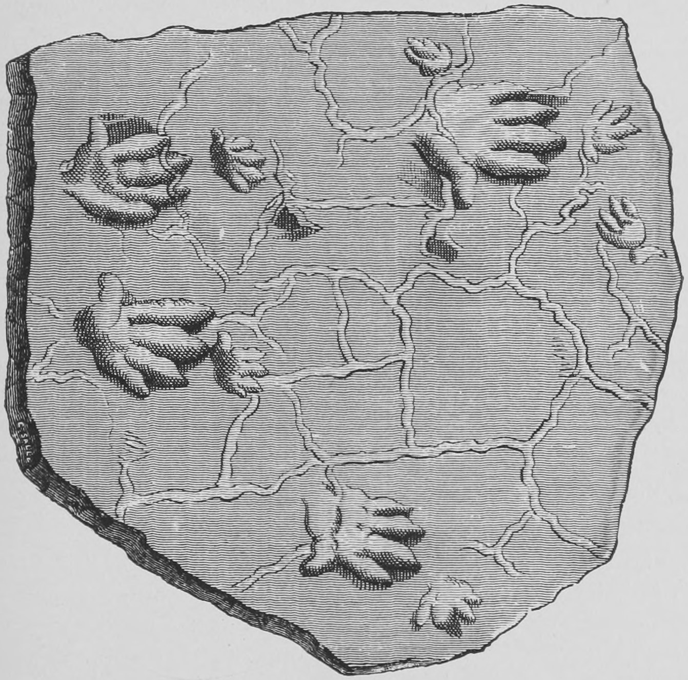
In Fig. 40 we have drawn a slab of Triassic rock, showing the five-fingered hand-like footprints of the Cheirotherium (as it was once called), a huge salamander-like animal.
TABULAR LIST OF THE GREAT GROUPS OR BRANCHES OF THE PEDIGREE OF ANIMALS
| I. | VERTEBRATA. (Back-boned Animals.) |
| Class 1. | MAMMALS. |
| 2. | BIRDS. |
| 3. | REPTILES. |
| 4. | AMPHIBIANS. |
| 5. | FISHES. |
| 6, 7 and 8. | Lancelets, Ascidians and Acorn-Worms. |
| II. | MOLLUSCS. (Mussels, Oysters, Clams, Snails, Slugs, Whelks and Cuttle-fish.) |
| III. | APPENDICULATES. (Insects, Crabs, Shrimps, Spiders, Scorpions, Centipedes, and Annulate Worms and Wheel-animalcules.) |
| IV. | ECHINODERMS. (Starfishes, Sea Urchins and Sea-Cucumbers.) |
| V. | FLAT WORMS. (Flukes, Tape-Worms, Water-flukes, etc.) |
| VI. | NEMERTINES. (Cord-like Sea-Worms.) |
| VII. | NEMATODS. (Parasitic Thread-Worms.) |
| VIII. | CORAL-POLYPS and SEA-ANEMONES. |
| IX. | HYDRA-POLYPS and JELLY FISH. |
| X. | SPONGES. |
| XI. | PROTOZOA. (Microscopic Unicellular Animalcules, Amœbæ, Gregarines, Flagellates, etc.) |
N.B.—The list does not contain the less important great groups, and is purposely made more simple than are the tables of classification used in scientific text-books.
TABULAR LIST OF THE CHIEF ORDERS OF THE VERTEBRATE CLASS
MAMMALS.
| Order 1 | Primates | Man, Apes and Monkeys. |
| ,, 2 | Insectivors | Hedgehogs, Shrews and Moles. |
| ,, 3 | Chiroptera | Bats. |
| ,, 4 | Carnivors | Dogs, Bears, Cats and the extinct Creodonts. |
| ,, 5 | Pinnipedes | Seals. |
| ,, 6 | Ungulates | Hoofed Animals: even-toed and odd-toed. |
| ,, 7 | Elephants | Elephants and their extinct ancestors. |
| ,, 8 | Amblypods | Dinoceras and Arsinöitherium. |
| ,, 9 | Toxodonts | Toxodon. |
| ,, 10 | Rodents | Rats, Rabbits, Beavers and Porcupines. |
| ,, 11 | Hyracoids | The Syrian and African "Coney." |
| ,, 12 | Sirenians | The Manatee, Dugong and Steller's Sea-Cow. |
| ,, 13 | Edentates | Sloths, Armadilloes, Ant-eaters. |
| ,, 14 | Cetaceans | Whales and Porpoises. |
| ,, 15 | Marsupials | Kangaroos, Opossums, Tasmanian Wolf. |
| ,, 16 | Cloacals | The Egg-laying Platypus and Echidna of Australia. |
N.B.—The list is not complete
TABULAR LIST OF THE CHIEF ORDERS OF THE VERTEBRATE CLASS
REPTILES.
| Order 1 | Dinosaurs | Huge extinct creatures, often as big as elephants. |
| ,, 2 | Crocodiles | Crocodiles and Alligators. |
| ,, 3 | Chelonians | Turtles and Tortoises. |
| ,, 4 | Lizards | Closely allied to one another, and having the epiderm moulded to form "scales." |
| ,, 5 | Snakes | |
| ,, 6 | Pterodactyls | Extinct: they had great flying wings supported by one finger. |
| ,, 7 | Theromorphs | Extinct: often with teeth resembling those of Mammals. |
| ,, 8 | Plesiosaurs | Extinct: swan-necked aquatic forms, with four paddles. |
| ,, 9 | Ichthyosaurs | Extinct: short-necked marine forms, closely representing among Reptiles, the Whales and Porpoises of the Mammalian series. |
N.B.—This does not profess to be a complete enumeration.
Chapter Two: Strata and Land Surfaces—Teeth and Bones—Extinct Men—Flint Implements—The Mammoth Elephants and Mastodon—Classification of Animals
Before giving you further accounts of extinct animals, I wish to point out to you that what I have to say is true, and not mere imagination.
Some people talk about the "fairy tales of science." There never was a more inappropriate phrase: it is altogether wrong to speak of fairy tales having anything to do with science. The wonderful things which science reveals to us are altogether remote from fairy tales, for in regard to the tales of science you can test what you are told, you can see the things of which I speak, you can ascertain the truth of what is asserted. That is the great pleasure of this study; one knows that the things one examines, however astounding and incredible they seem, really exist, and are not mere imagination or fancy.
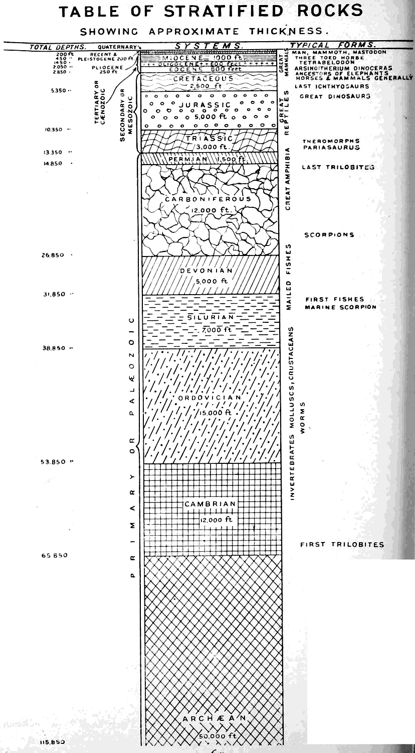
I want now to refer to these large diagrams (Figs. 41 and 42). Fig. 41 should be carefully examined. It represents what has been discovered with regard to the succession of deposits, those stratified deposits of which I spoke in my last lecture. On the left-hand side is stated the thickness of each deposit, so far as it has been ascertained.
Most of the extinct animals, all the great extinct animals I have to speak about, come within the upper part. We have an enormous thickness of stratified rock beneath, which contains only marine things, fishes, a few crustaceans, and things of that kind. But all the more interesting great animals have left their bones in the higher strata. The uppermost layer (the recent and Pleistocene) is only some 200 feet in thickness, yet it indicates a period of something like 500,000 years. This being so, you can judge by the thickness of subjacent deposits what an immense lapse of time is represented. Before we get to the chalk we get down nearly 3,000 feet. The thickness of the chalk itself is another 2,500 feet.
The estimate thus given probably does not fully represent the time which has elapsed. If you take a thousand years for each foot, you only get an approximate measure of the time represented, because a great deal more time has passed than is actually shown by the permanent deposits or strata. Strata have been broken up by the sea and water, and have been deposited again and again; and it is probable that a much longer time has elapsed than one thousand years for each foot of the deposits which form the stratified crust of the earth.
An important general fact, which I cannot dwell on further, is that whilst it is true that the great animals occur in the later stage of the world's history, there is a gradual succession from simpler to more complex forms of life. We get fishes at the top of the Silurian; and we get in the Carboniferous great amphibians; and the first reptiles in the Permian; and then we get birds and crocodiles in the Triassic; and the first hairy warm-blooded quadrupeds in the Jurassic. Thus the different kinds of animals succeed one another in the order of increasing complexity of structure so that the highest animals are the latest to appear.
This map of the world (Fig. 42) has special interest, for it shows the present position of different kinds of animals. It is meant to show something of the history of the migrations or movements over the surface of the earth of the large animals which have lived upon it. The line which separates New Zealand from the rest of the world, which we call Theriogæa—the land of big animals—shows that the large islands of New Zealand have no such animals upon them. Till man went there some thousand years ago there were no large animals. The largest animals were great birds. There were no cattle, or cats and dogs—not even mice. Thus this piece of land seems to be separated from the history of the movement of animals in the rest of the world. It is an old and detached land-surface. Then you will see a second line between Australia and the rest of the world.
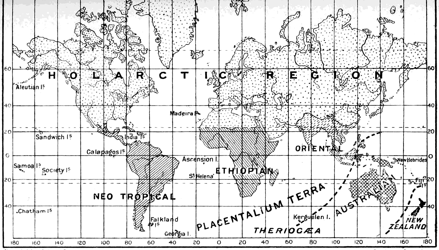
Australia is distinguished by its marsupials (kangaroos, wombats, phalangers, etc.). The young of the marsupials are very small when born and are placed by their mother in a pouch of skin overlying her teats. Those animals which are nourished inside the mother before they are born are of a much larger size at birth. They are the Placentals, and there are no aboriginal Placentals in Australia.
The greater part of the world (the rest when Australia is cut off) may be divided into the great Holarctic surface, the northern strip which comprises North America, Europe and the Northern part of Asia, while projecting downwards are three other regions, South America or the Neo-tropical, the great African or Ethiopian region, and the Oriental or Indian region. The animals of which fossil remains are found in the Holarctic region have migrated into these projections of the land which subsequently became of their present shape, at different times in the world's history.
The Neo-tropical region of South America was at one time a separate mass of land, and upon it lived very peculiar animals, such as the great sloths and armadilloes, and strange birds.
In the Holarctic region we find, either still living or buried in recent strata, the great hairy mammals, elephants and cattle, antelopes, deer, camels, horses, rhinoceroses, tapirs, pigs, hippopotami, tigers and lions, and such forms. When we dig down only to the depth of a few feet, in river gravels and comparatively modern deposits, we find all the big creatures in this region as shown by their fossilized bones. But, owing to some change of climate and other conditions not very clear, most of them left this region and migrated to the southern projecting regions.
One of the most curious results of this emigration is that at the present day the tapir is found alive in the island of Sumatra and that it is found alive also in Central America. At one time naturalists were much surprised to find a tapir in the new world like the tapir in the old world, and nowhere else but in these limited spots, remote from each other. But now we know that tapirs existed all over the Holarctic region, for we find there their fossil remains; we recognize them by the shape of their teeth and bones which we dig up. Even in England, in Suffolk, we find the tapir in the deposit known as the Red Crag, and again in different parts of Germany, France and Greece, and even in China and in North America, tapirs are found buried in the sands of Pliocene and Miocene age. The present race of tapirs existing in the East Indies and in Central America are as it were the outlying survivors of those which existed formerly all over the great Holarctic region.
Such facts as these about the tapir indicate the importance of knowing where particular fossil animals are found; for thus we are enabled to come to some conclusion as to the former connexion of different land surfaces of the world with one another.
The question must have occurred to many of you,—How do we recognize fragments of bones found in the earth? How do I know that a fragment I may find is the lower jaw of a creature like the horse? or that bones I may dig up are the bones of a tapir? How do I know that a given skull is that of a reptile? and that a given shell was inhabited by a creature like the nautilus?
We are able to know these and like matters because the shape of different parts of each kind of animal is very constant. The kinds which are like one another in other respects are like one another in the details of their bones and teeth, even in such minute points as the microscopic texture of the bones. An immense mass of facts about such things is known, and when set out in orderly fashion is termed the science of comparative anatomy or animal morphography.
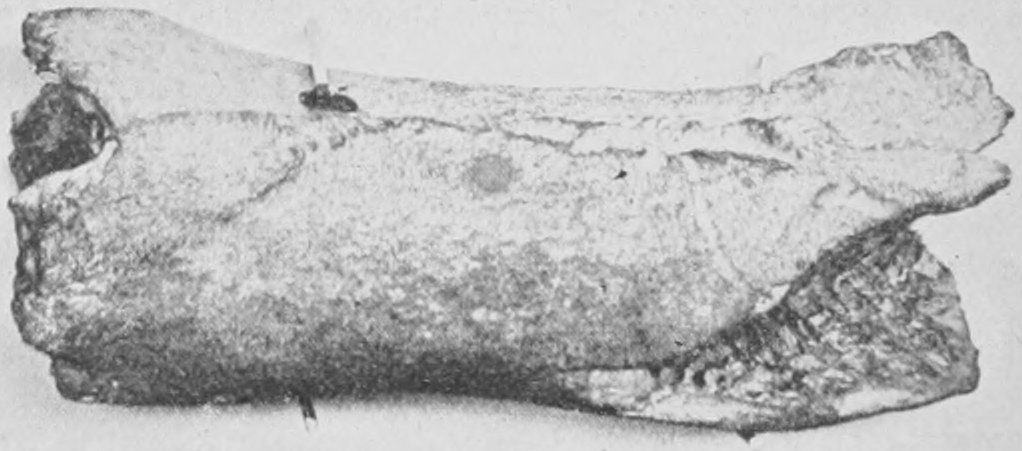
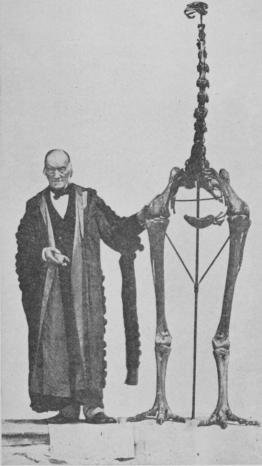
The first photograph I have to show in this chapter is of a piece of bone which was sent fifty years ago to Professor Owen by a gentleman in New Zealand who had lately arrived there, and who had found it in his garden. Professor Owen, on examination, was able to say from the general make and structure of the bone that it was the bone of a bird. It was about seven or eight inches long (Fig. 43). On examining the ridges and various marks on the bones, Owen was able, from his knowledge of the character of bones, to say that it was identical with the middle part—the ends were broken off—of the thigh bone of an ostrich. He ventured then to publish that this bone was a proof that there existed formerly in New Zealand a huge terrestrial bird like the ostrich, only bigger. After a few years, more bones were sent to Owen from New Zealand, which entirely confirmed what he had said: and in the course of a few years he was able to put together from the bones sent a skeleton with enormous legs and neck, the skeleton of the ostrich-like bird the Moa of New Zealand. In Fig. 44 you see Professor Owen himself at the side of the restored skeleton. Since that time a great number of these birds have been found buried in the morasses and comparatively recent deposits of New Zealand, showing that many of them existed alive some five or six hundred years ago, and that they were then probably hunted out of existence by the ancestors of the present Maoris. I shall have a few more words to say about the giant birds of New Zealand in a later chapter.
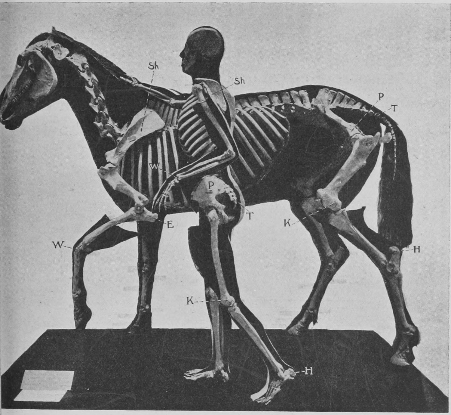
In Fig. 45 we have the photograph of a very fine preparation in the Natural History Museum, showing the skeleton of a man and a horse side by side. The main object of this comparison is to show that, though so different in general bearing and form, all the bones of a man correspond in detail with those of the horse. The thigh bone of the horse and the thigh bone of the man, the knee (called the "stifle") of the horse and the knee of the man, correspond. The man has a short foot, the horse a long one. The upstanding bit at the back of the horse's leg called the "hock" is really the heel, and corresponds to the heel bone which you can distinguish in the man's skeleton. So also the fore-arm and shoulder-blade correspond in the two skeletons.
Accordingly, as animals are alike or unlike in the details of their structure, so we can group them into divisions and sub-divisions (see the list of classes at the end of Chapter I). There are certain marks by which it is easy to recognize fragments of bone, dug it may be out of a quarry or railway cutting, and to know at once the division or kind of animals to which the owner of the fragments belonged. I have already alluded to the fact that the strata of the earth are revealed to us by cliffs on the sea-shore, by exposed rocks and by river banks; and I would add by such activities of man as the digging of quarries and railway cuttings. Suppose that you find a skull in such a digging—there are marks by which you can tell whether it belongs to a mammal or reptile.
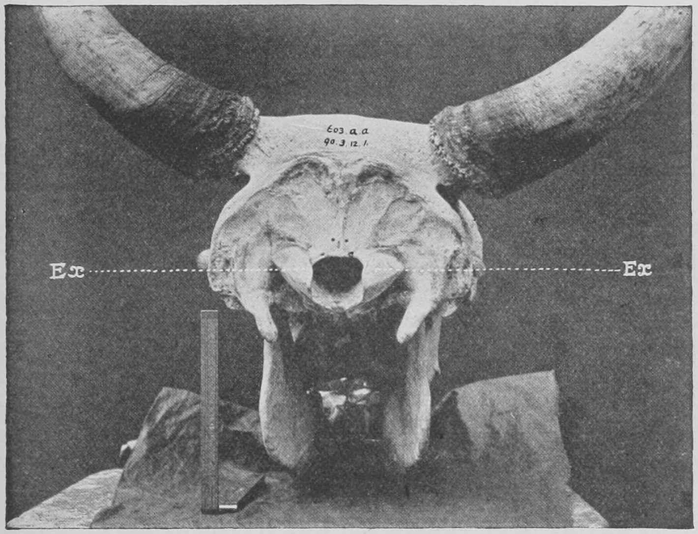
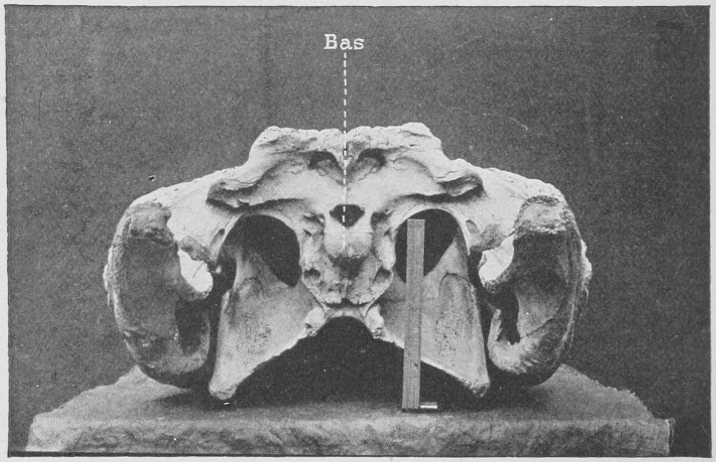
In Fig. 46 I have photographed the whole back part of a skull which contained the brain, and you see where the spinal cord entered the skull to join the brain. In this creature (an ox) there are two bony surfaces (marked Ex, Ex) forming the joints or condyles of the skull by which the first neck-bone or vertebra was fastened to it whilst allowing a rotating movement. All mammals' skulls are provided with this pair of knobs or "condyles." But in the crocodile's skull (Fig. 47) you will see below the aperture for the spinal cord only one large condyle (marked Bas). From such a fragment of the skull then you can at once tell whether to place the creature to which it belonged among the hairy warm-blooded quadrupeds called mammals, or with the reptiles. A bird is like a reptile in having a single joint or knob at the back of the skull.
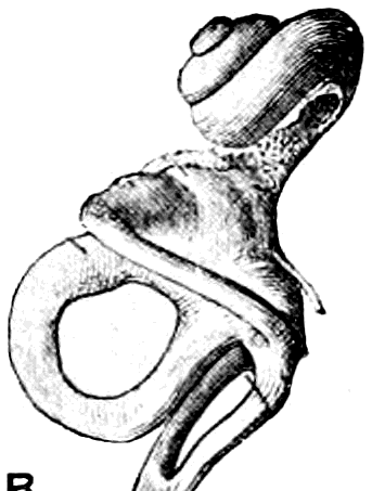
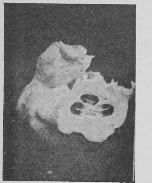
As an example of the definite marks by which bones can be referred to their proper classes, the following is a curious point. Fig. 48 is a drawing of the internal ear of man—the soft part of the ear inside, embedded in bone. It consists of three loop-like canals and a snail-like coil. All hairy mammals have that snail-like construction of the internal ear. In Fig. 49 is photographed the ear-bone of a mammal's skull cut through, and you can see the place for the snail-like soft ear—the cochlea or internal spiral of the ear, as it is called. No other animals except the mammals are known to possess a spiral internal ear, and all known mammals do possess it. If, therefore, you discovered a fragment of bone showing this spiral-like space you would know that the bit of bone must in all probability belong to a mammal.
At the beginning of the nineteenth century a portion of a great elongated skull was brought from America to Europe, dug out of the sands of Florida. It was thought to belong to a reptile like the crocodile, and was called Basilosaurus. But the naturalist in whose care it was, on showing the specimen to a friend (Herman von Meyer) dropped it on the stone floor of his museum and cracked the back of the skull. The crack exposed the spiral cavity or cochlea of the ear, and thus it was shown that the specimen was the skull of a mammal. Sure enough, it turned out later to be the skull of a kind of whale (Zeuglodon).
Teeth are of great help and importance in determining the sort of animal to which a fragment belongs.
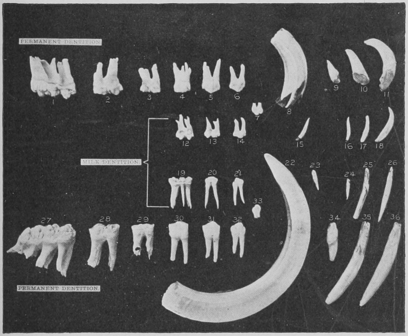
Fig. 50 is a photograph from a specimen prepared in the Natural History Museum. The wild boar or pig occupies in regard to teeth a sort of central position among mammals (hairy warm-blooded quadrupeds). Its teeth are such that to them you can refer, as to a standard pattern, the teeth of all other mammals. There are three middle teeth in front in the upper and lower jaw, chisel-like teeth, the incisors. Beyond these are the great canine teeth: then the cheek teeth follow. These are seven in number, four in front which are replaced—that means that second teeth come to take the place of the first—and three hinder ones, which are never replaced. If you look at the surface of these cheek teeth you will find they are broad, with many tubercles, fitted for grinding great varieties of food. There are seven of these cheek teeth on each side in each jaw, upper and lower, one canine, and three incisors, so that eleven on each side in upper and in lower jaw or forty-four teeth in all is the complete number, the typical number—the most characteristic number in the group of hairy mammals. Many have less, but among the immediate ancestors of those mammals with "reduced dentition" we find a larger number of teeth, and in their remote ancestors the complete typical number is discovered.
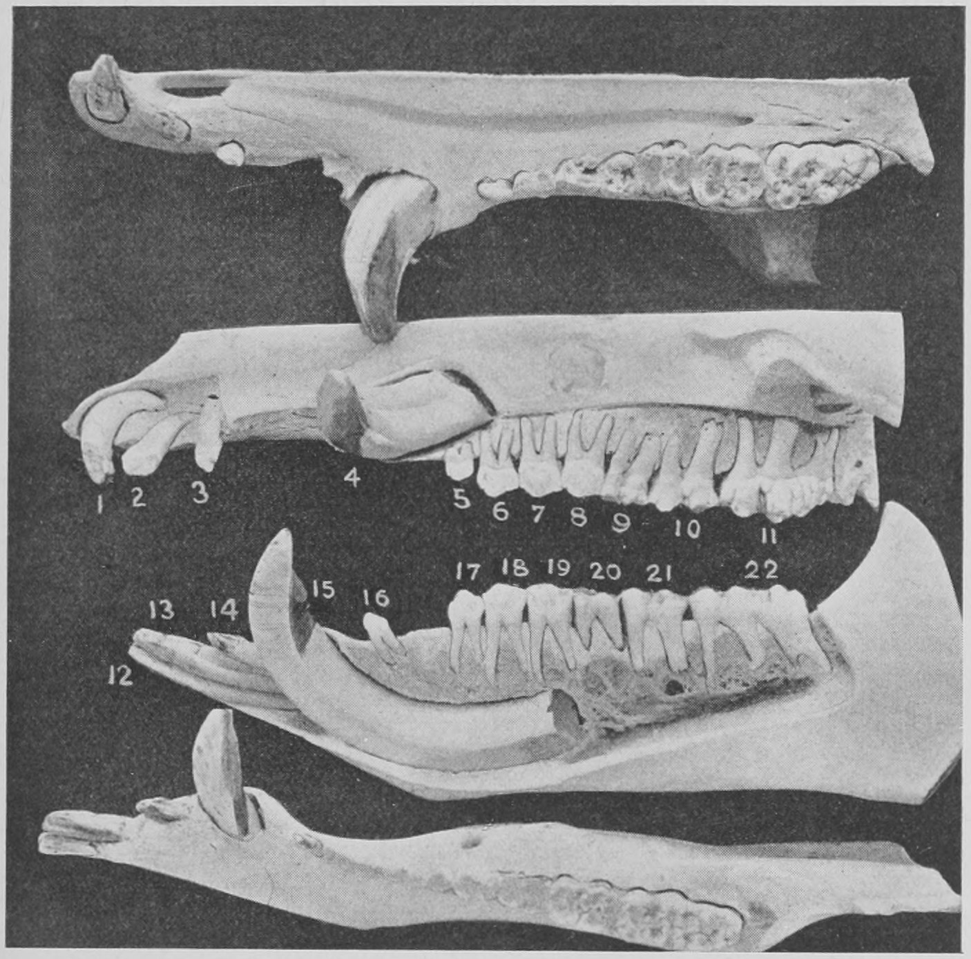
It is important to notice that whereas the front teeth have a single fang by which they are implanted in the jaw the cheek teeth have two fangs, as shown in Fig. 51. Teeth with two fangs appear to be peculiar to mammals. Other animals have only single fangs to all their teeth, as mammals have for their incisors and canines (as a rule).
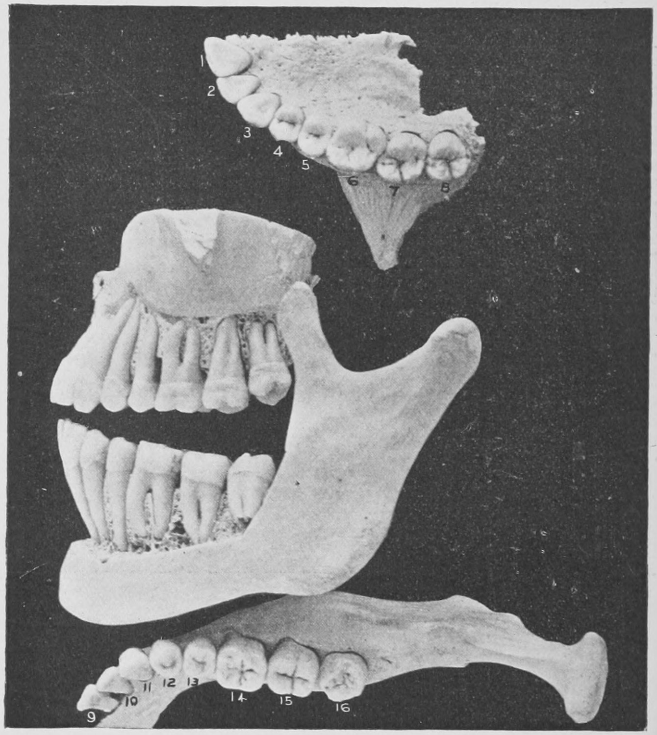
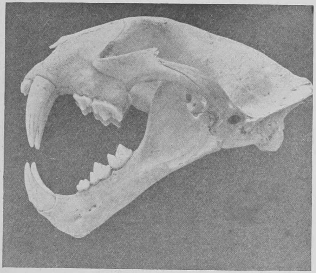
The human teeth (Fig. 52) are reduced in number. There are only two incisors above and below on each side; then the small canine or dog-teeth, one on each side; then five cheek teeth or "molars," two smaller and three bigger. From a single tooth we could tell whether a piece of jaw-bone belonged to a man or not. Though like a monkey's, a man's tooth can be distinguished from it and from all other teeth. In the next figures we have photographs showing certain modifications in the teeth of mammals. You see in the Clouded Tiger (Fig. 53) that the teeth are few in number, and are sharp, for cutting or tearing flesh, whilst the canine teeth are very large.
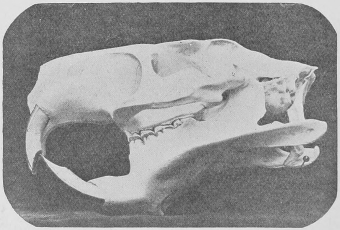
In Fig. 54 the skull of a great rat, as big as a beaver or fair-sized dog, is photographed. The front teeth (only one on each side above and below) are chisel-like, and very large, to enable the rat to gnaw wood.
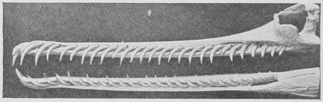
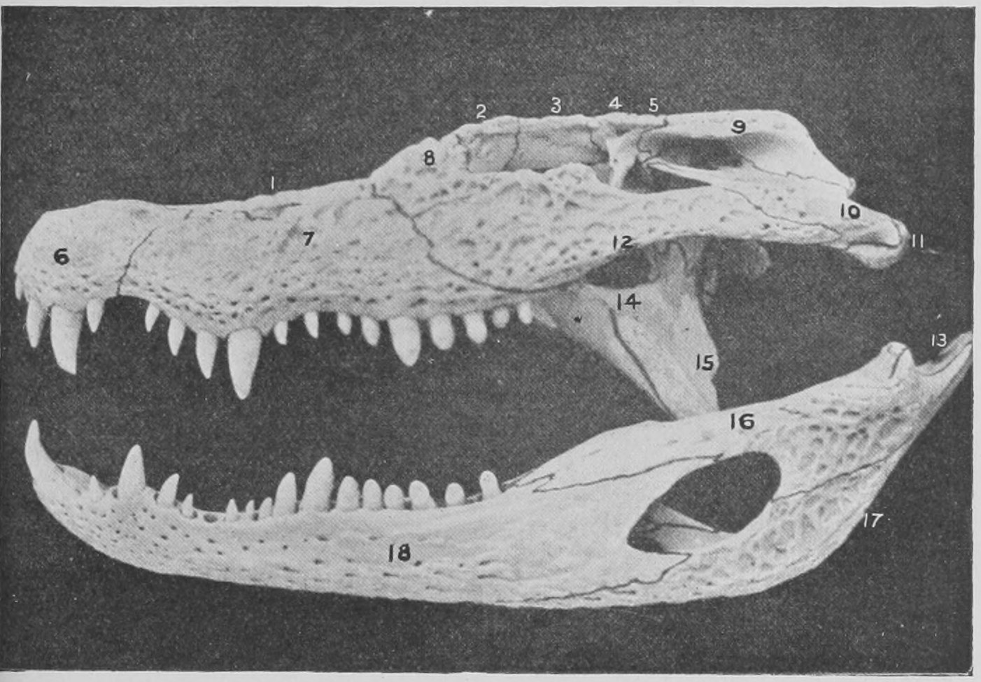
In reptiles you no longer get complex cheek teeth. All the teeth are peg-like. They have no grinding teeth with big surfaces, and all the teeth have a single fang (Figs. 55 and 56).
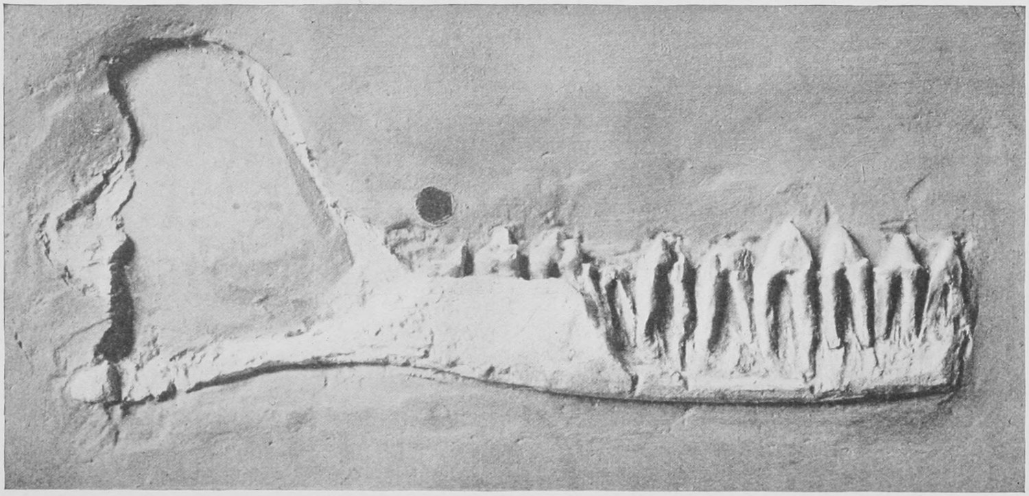
The fossil jaw shown in Fig. 57 came from Stonesfield in Oxfordshire. It is embedded in hard Jurassic slate, and is one of the most ancient evidences of the existence of a mammal. The sight of its double fangs at once rendered it almost certain that the teeth must be those of a mammal: the whole shape of the jaw is like that of a small mammal, such as the hedge-hog.
We must now take up again the general story of extinct animals; and to do so we will first of all go back, so to speak, a little way into the strata deposited on the earth's surface—just far enough to take us beyond the range of written history or record, which barely reaches further than four thousand years.
Recent explorations in ancient cities, Egypt and other parts of the East have brought out from layer after layer of rubbish and mud the different remains of man, different instruments, utensils and works of art. As one gets deeper one finds remains showing different habits and ways of life. But all are practically within the historic period. Beyond that we come to a period of which there is no tradition or written record, but of which we have evidence only by the remains we find; flint instruments, carvings, and even occasionally some human bones. The most important of the prehistoric remains of man take us back, to judge from the position in which they are found, some 150,000 years. These are the remains, found in river gravels in England and France and other countries, proving that man lived here in a savage state with the Mammoth, the Rhinoceros, Hyena, Cave Lion and Cave Bear.
Fig. 58 shows two flint implements which these men manufactured and used. A flint instrument of this kind was found more than a hundred years ago in the gravel in Gray's Inn Lane, in London, and was figured and described; but its great antiquity was not recognized at that time.
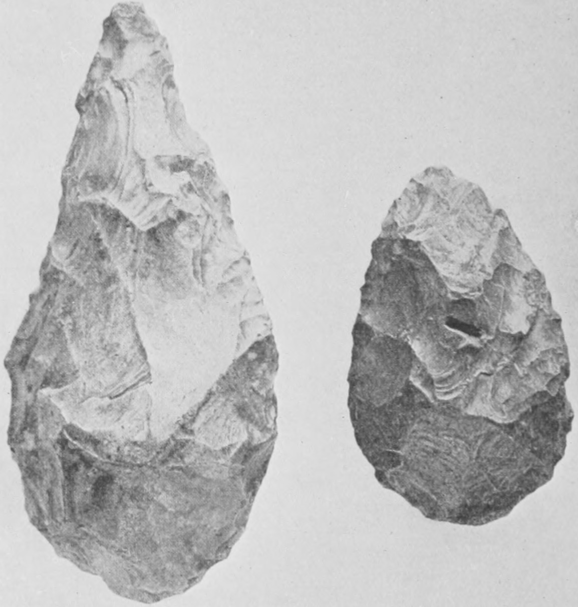
In the middle of the last century, attention was drawn to these flint instruments found in the gravel of the river Somme by a French antiquarian, M. Boucher de Perthes. He got immense quantities of these worked flints from the neighbourhood of Abbeville and Amiens, and he maintained they were the work of men. They were clearly, from the depth of gravel under which they were found, of enormous antiquity. The matter was gone into carefully at the time; geologists and naturalists took keen interest in it, and the great antiquity of man in Europe was established. And besides these implements in the gravel others have been found in caves associated, as in the gravel, with the remains of animals which have long ceased to exist in this part of the world. These are such mammals as the reindeer, the hairy rhinoceros, the great Irish stag, the cave bear, the cave hyena and the lion. Huge wild cattle, such as the Aurochs or Urus of Caesar, and the Bison, existed then in quantity. In some places the actual bones and skulls of these primitive men have been found with the bones of extinct animals.
The skulls of primitive men and of modern men show a certain difference in shape. If we take two skulls, that of a man and a monkey (Fig. 59), and draw a line from the region just over the nose, between the ridges of the brow, and run it back to the occipital ridge at the back of the skull, there is left above the line a great hemispherical dome in the human skull, whereas in the monkey the space left above is much flatter, much shallower.
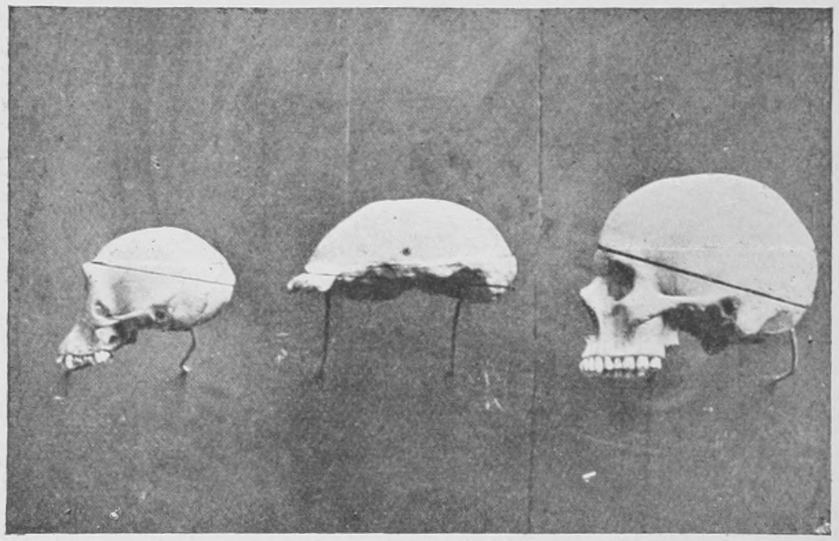
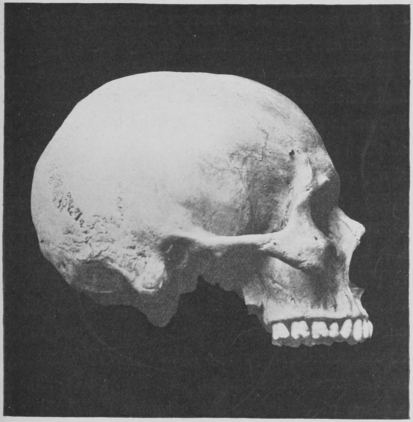
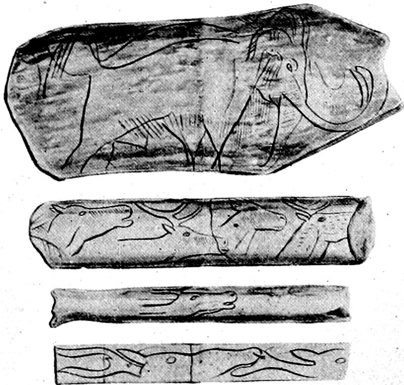
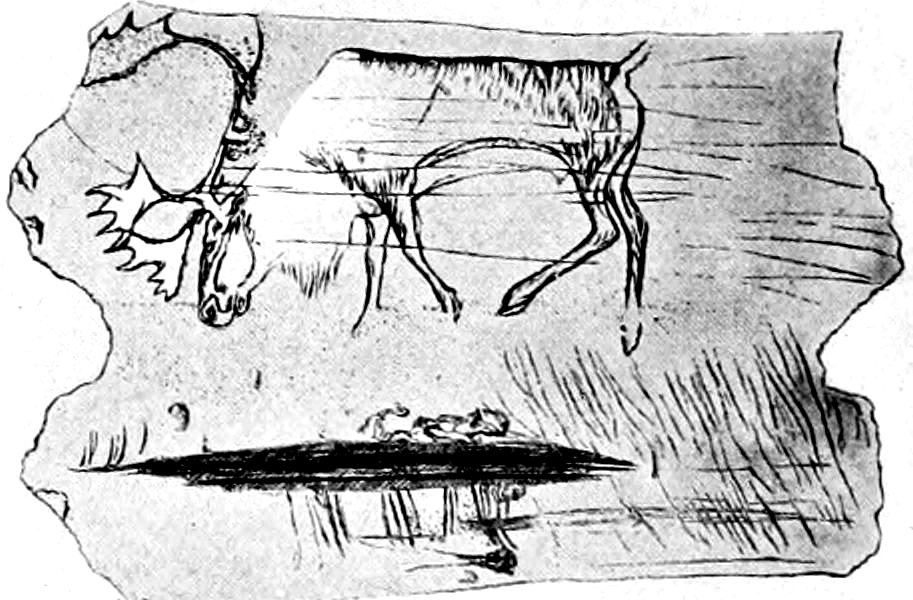
In a river gravel in Java the imperfect skull of the so-called Pithecanthropus, or monkey-man, was lately discovered. It is really, in its main features, a human skull. A photograph of it is seen in Fig. 59—the middle one of the three figures. It has a shallow upper region, much like that of a monkey. Other shallow skulls of primitive men have been found in caverns, such as those of Spy in Belgium and in the sand of the Neanderthal on the Rhine. It seems certain that primitive man had a shallower brain than the more recent man. But some of the prehistoric men seem to have been able to draw, and to have exhibited great skill in that art. It is difficult to say whether there was more than one race present at this time, and whether the men of the shallow skulls were the same men who made the drawings. In one of the caves of France inhabited by prehistoric men, and thickly strewn with their chipped flints and with the bones of extinct animals eaten by the men, a piece of a mammoth's tusk has been found with a mammoth carved upon it (Fig. 60) evidently by the men who lived there. We also find the heads of reindeer, carved upon pieces of bone. The photographs reproduced in Figs. 60 and 61 are from drawings of the actual specimens. In Fig. 61 is shown a piece of an antler upon which a reindeer is cleverly outlined. The tuft of hair below the chin is shown, and the great feet and the extra toes are correctly pictured. Clearly the men who drew this reindeer lived with the reindeer. And besides the reindeer, living with those men in the South of France was the great mammoth.
The mammoth was like an Indian elephant, but with a coarse hairy pelt and its tusks had a slightly different curvature from that seen in the Indian elephant. It was a little bigger than the biggest Indian elephant.
The mammoth has left its remains all over the Holarctic region. Even in our own country we are continually coming across tusks and teeth. In the Natural History Museum there is a whole skull, with enormous tusks, which was dug up in a brick-field at Ilford in East London about twenty-five years ago. From this brick-field I used to get many remains of mammoth, rhinoceros and hippopotamus when I was a boy. When this mammoth's skull was found by the workmen, the authorities of the British Museum of that day sent down a man to remove the specimen with the greatest care, and it is now at the Natural History Museum.
The remains of the mammoth are found in enormous abundance throughout the Holarctic region. It is probable that this huge beast existed later in Asia and Siberia than in our part of the world. In the north of Siberia complete carcases of the mammoth and also of the hairy rhinoceroses are found in a frozen condition, with the skin, hair, trunk and soft parts complete.
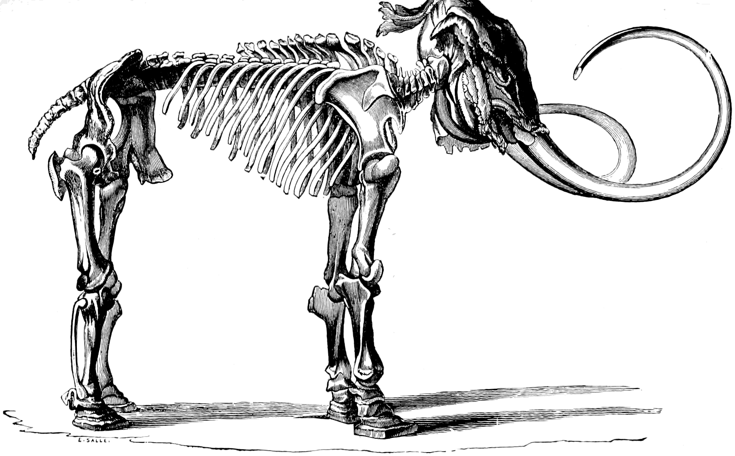
At the beginning of the last century one of these frozen carcases was removed to the Museum at St. Petersburg. It is from this specimen, drawn in Fig. 62, that we know that the mammoth had a hairy skin.
It is an interesting fact that the newborn young, both of the Indian and the African elephant have a complete coat of fairly long hair, which disappears in a few weeks. So the mammoth is not really peculiar in this matter.
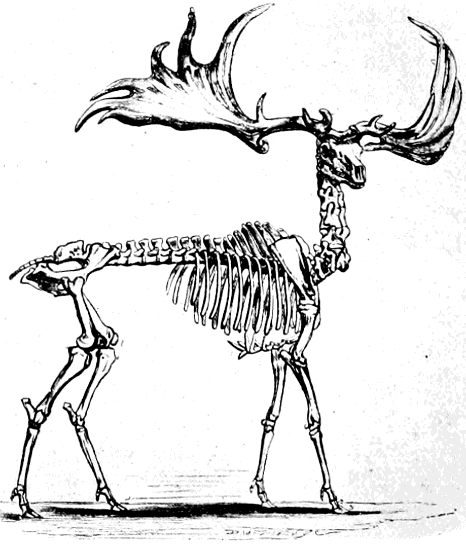
In Fig. 63 is shown the skeleton of the largest and most beautiful of all the deer tribe; it is now extinct, but existed later in Ireland than anywhere else, and in great numbers. The bones are found in the moss and bogs of Ireland.
It was co-existent with primitive man, and perhaps survived in Ireland till nearly historic times. Why it died out there is a difficult thing to explain.
As our explorations into the river gravels of only twenty or thirty feet depth have brought us into contact with the mammoth, I propose now to say something more about recent and extinct elephants, and to take a glance at the past history of the elephant tribe.
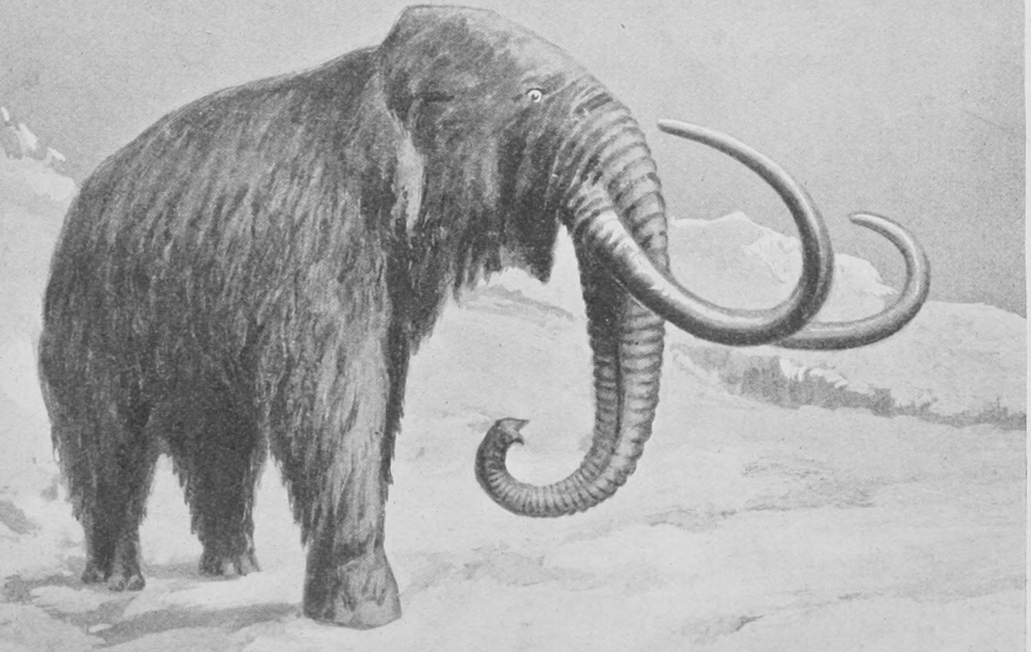
Fig. 64 gives a careful restoration of the hairy mammoth as it must have appeared in life, and in Fig. 65 we have a photograph from life of the Indian elephant. In the Indian elephant you should note the comparatively small ear and the high forehead.
Fig. 66 is a photograph from life of the African elephant. It has a longer head and much larger ear than the Indian species.
The biggest Indian elephant is very rarely as much as eleven feet high at the shoulders. But some specimens of the African elephant must grow to twelve and possibly thirteen feet in height, if we may judge by the size of their tusks.
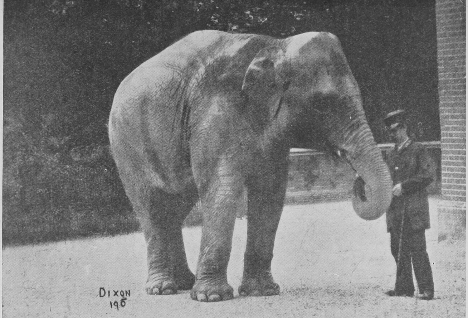
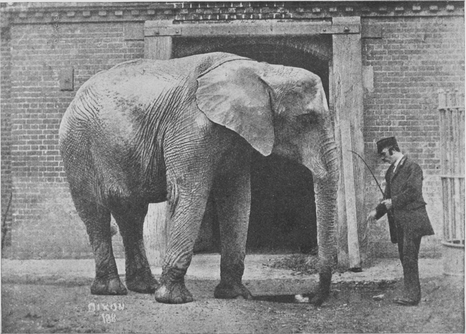
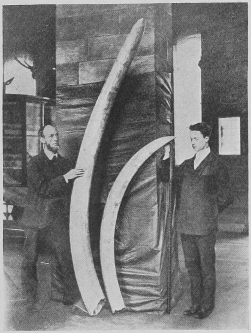
Fig. 67 shows two elephants' tusks: the two specimens are in the Natural History Museum.
One is a very fine Indian elephant's tusk, weighing seventy-two pounds—as big an Indian elephant's tusk as is to be found in any museum. The other is an African elephant's tusk; it is ten feet two inches in length, and weighs 228 pounds. It is the biggest tusk ever seen in the ivory market, and was purchased for the Museum in 1900. Elephants' tusks have for many years been imported from Africa, and never has one been seen to approach this in size. Anything near 150 pounds in weight is considered enormous; and this weighs 228 pounds. It was brought from Zanzibar ten years ago, and is one of a pair which probably formed part of the treasure collected by Emin Pasha.
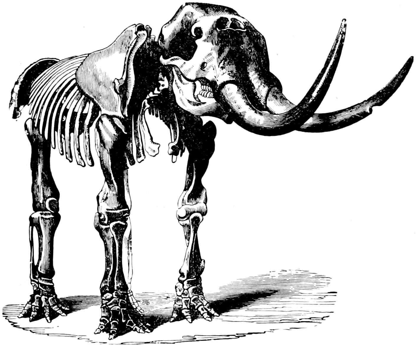
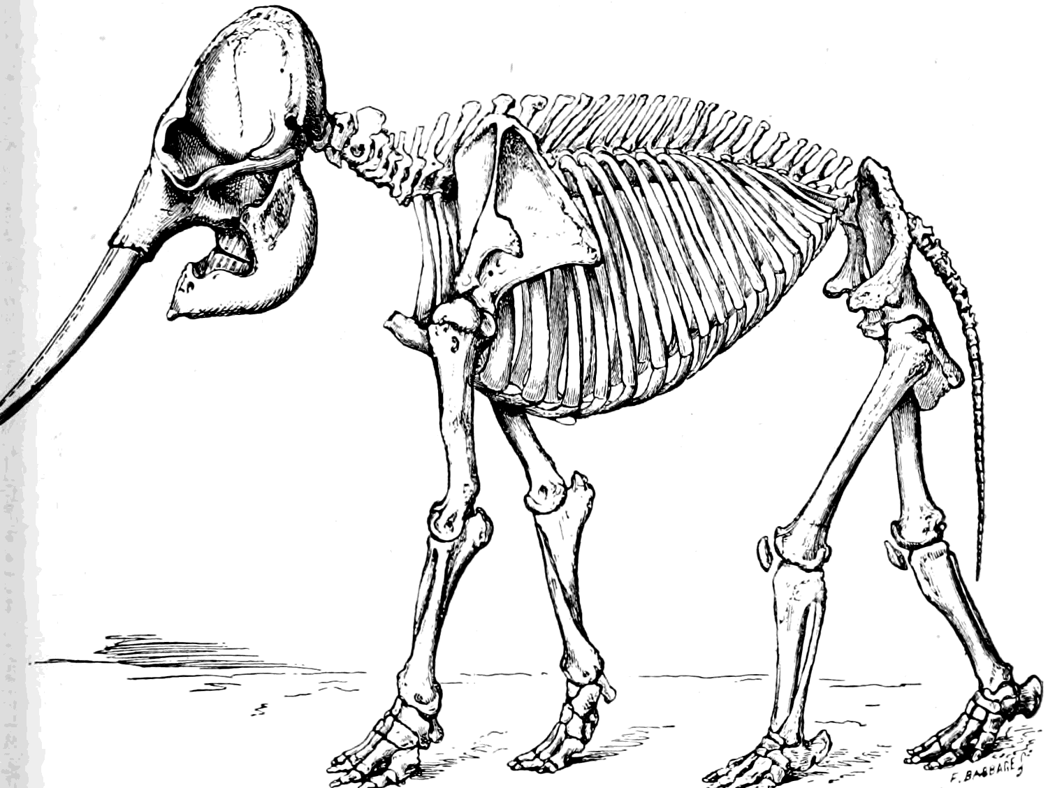
In Fig. 68 we have represented the skeleton of a creature very similar to the true Elephant, but that it has slightly different teeth and a more "snouty" or elongated head and jaw. It is the American mastodon. The skeleton should be compared with that of the mammoth given in Fig. 62, and with that of the Indian elephant shown in Fig. 69.
In the United States (Ohio and other localities) very complete remains of this enormous creature have been found in bogs and morasses which are probably not more ancient than the peat bogs of Ireland in which the great Irish stag is found. Man was certainly contemporary with some of the American species of mastodon. But in Europe no mastodons survived to so late a period. Other and older species of mastodon seem to have preceded the elephants in Europe, Africa and Asia, and in fact to have been the ancestors from which elephants were derived.
Chapter Three: The Ancestral History of Elephants—Extinct Horses and Rhinoceroses—The Arsinoitherium
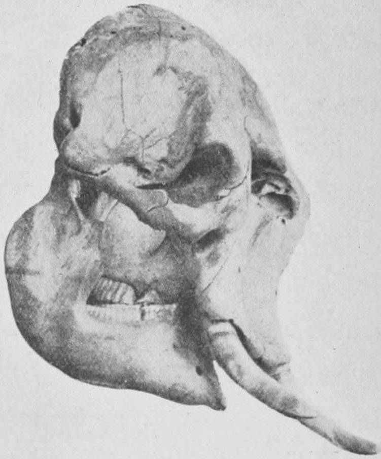
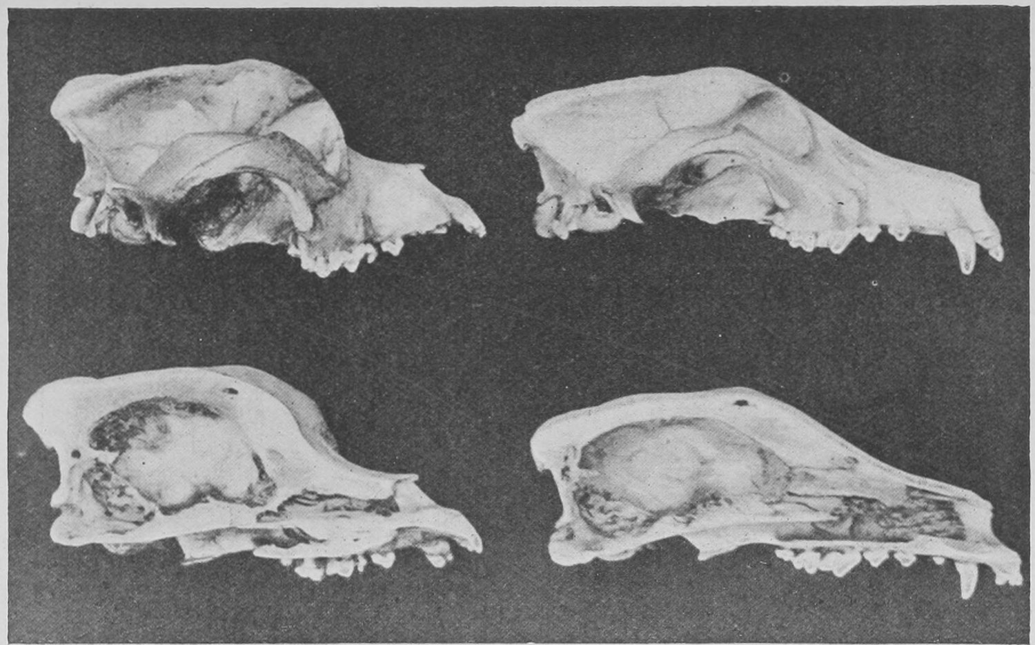
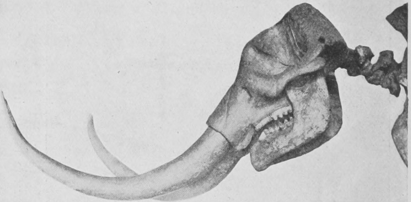
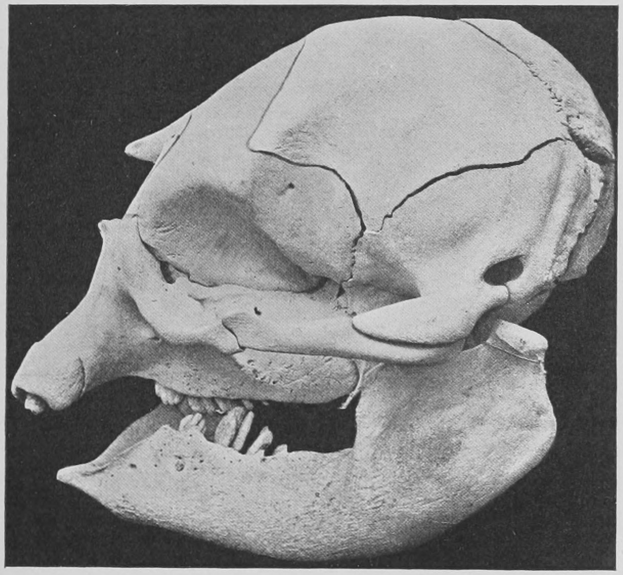
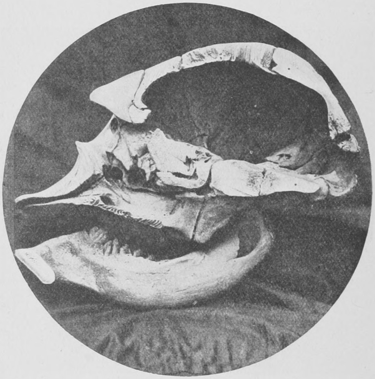
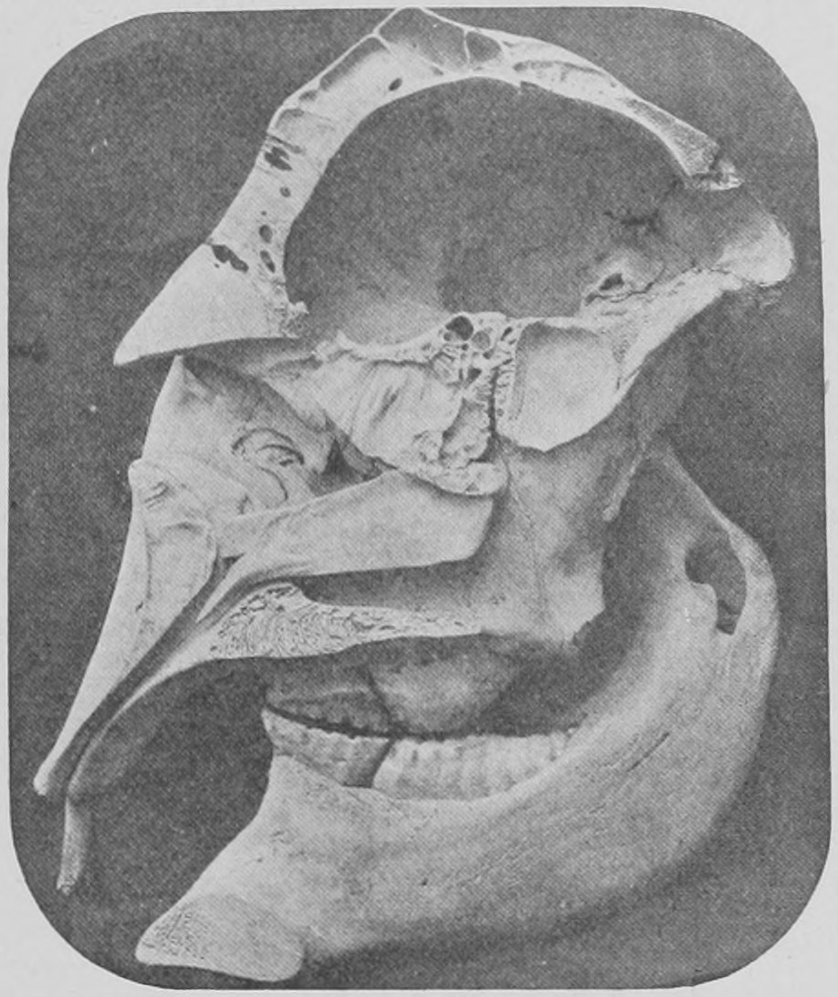
When we compare the American mastodon with true elephants, viz., the African, the Indian, and the Holarctic one called the mammoth, we find in the first place that though the mastodon is as big a beast as any of these, and very close to them in the form and arrangement of its bones, yet there are two important differences to be observed. These relate first to the shape of the head, and secondly to the shape and number of the teeth. If you look at an elephant's skull (Fig. 70) and compare it with that of a dog (Fig. 71) or pig, you will be struck by the abrupt way in which the bones of the face are set. The face is almost straight so far as the bony parts are concerned—both the upper and lower jaw are quite short. There is, in fact, no "snout" indicated in the bony skull. This extreme shortening or pushing in (as it were) of the face is similar to what occurs in bulldogs and pugs as compared with ordinary long-snouted dogs, as is shown in Fig. 71, only in the elephant it is carried further than in any dog. We may call this shortening of the face "bull-dogging." This "bull-dogging" of the skull occurs in a South American race of cattle called the Neatta breed. It seems that the ancestral elephants must have had a long face and bony snout like other animals, but that their descendants have gradually become "bull-dogged." The mastodon's skull (Fig. 72) shows far more of a projecting bony face or snout than does that of the elephant, and this would lead us to suppose that the mastodons are more primitive, that is to say, more like the ancient ancestors of elephants, than are the true elephants. An interesting fact in this connexion is that the young new-born elephant has a more "snouty" skull than the grown-up elephant, as is shown by Figs. 73, 74 and 75. It is often the case that very young animals show features in which they resemble their ancestors, which disappear as the young creatures grow to full size.
It is not only in having a more elongated face that the American mastodon is of a more primitive build than the true elephants. Its teeth also are less peculiar than those of true elephants and more like in number and shape to those of the ordinary, more central kinds of mammals, such as the pig (see Fig. 50 in the last lecture). The elephant has two enormous incisor teeth in the upper jaw, in front—the tusks. There are no corresponding teeth in the lower jaw. Then there is a gap in the series, and we come to the cheek teeth, which are very strange. The jaws, both upper and lower, are so short, and the teeth are so big, that there is only room for one tooth or a tooth and a half on each side above and below at one time. An elephant only ever has three full-sized cheek-teeth on each side above and below (twelve in all), and these push from behind forwards—the first getting worn out and pushed forwards as the second comes forwards, and this again wearing out and disappearing as the third pushes itself into place from the back of the jaw. Three little "milk teeth" or first-teeth of the molar series precede these on each side above and below, and are lost one after the other—between the second and fifteenth years of life. The first big molar comes into place in the fifteenth year, and lasts for ten years, when its place is taken by the second, which is already showing its crown in the twentieth year. The third comes forward in the same way about twenty years later.
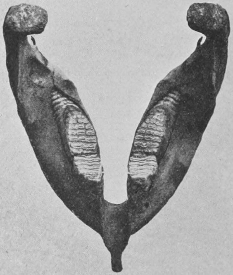
The molar teeth of the Indian elephant and of the mammoth have a great number of narrow transverse ridges set across the crown of the tooth. As many as twenty-seven of these ridges are seen on the biggest molar tooth when it is in place, and the whole surface is worn by grinding. In Fig. 76 the ridges on the teeth are shown, but not to the full number, as the front tooth is reduced in size by wear, and the hinder one has not yet got all its crown into play. In Fig. 77 is shown a photograph of the lower jaw of an African elephant. Only one big molar tooth on each side is in position, and it has eleven transverse ridges. This is the most the African elephant ever has. It will be seen, by comparing the figures, that the ridges of the African elephant are much wider than those of the Indian. The corresponding tooth of the Indian elephant, owing to the narrower shape of the ridges, would have twenty-seven of them in view when fully "cut." Now there is no doubt that the increase in the number of the ridges and their narrow form is a late and special character of the elephants. Their cheek-teeth would be more like those of pigs, tapirs and bears, if they had fewer transverse ridges. Accordingly, in correspondence with the view that the mastodons are more primitive in their characters than the true elephants, we find that their cheek-teeth have very few transverse ridges—from two to five (fig. 78)—and that the jaw is relatively longer, so that there is room, not only for two complete crowns of molars to be in position on each side at the same time, but even for three. Thus we approach nearer to the central or "typical" condition of the mammals' teeth which, as we have seen in the pig (fig. 50) shows seven cheek-teeth in position on each side in each jaw at once—of which the front ones are second-teeth and were preceded by milk-teeth—whilst the three big back ones are not preceded. In tracing the ancestry of living mammals through extinct ancestors of different succeeding geological ages, we expect to find even the strangest and most curiously modified creatures, such as are the elephants in regard to their teeth and jaws and the horses in regard to their toes—preceded by forms which bring us nearer and nearer, as we recede into the past, to a sort of common form or "type" of the mammalian group—a hairy-coated creature, with five toes on each foot, the typical dentition or tooth series of three incisors, one canine, four front or fore-molars, and three back molars on each side of each jaw, with three or four tubercles or knobs on the crowns of the molar teeth. And we do not expect this remote ancestor to be very big—not much bigger than a dog—since great size is a peculiarity implying long and special predominance.
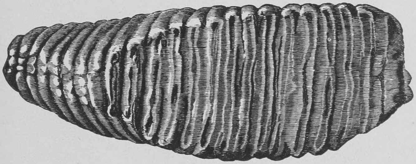
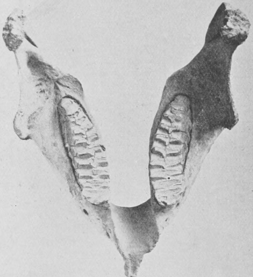
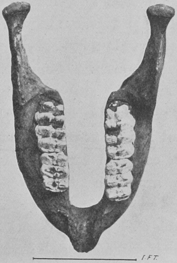
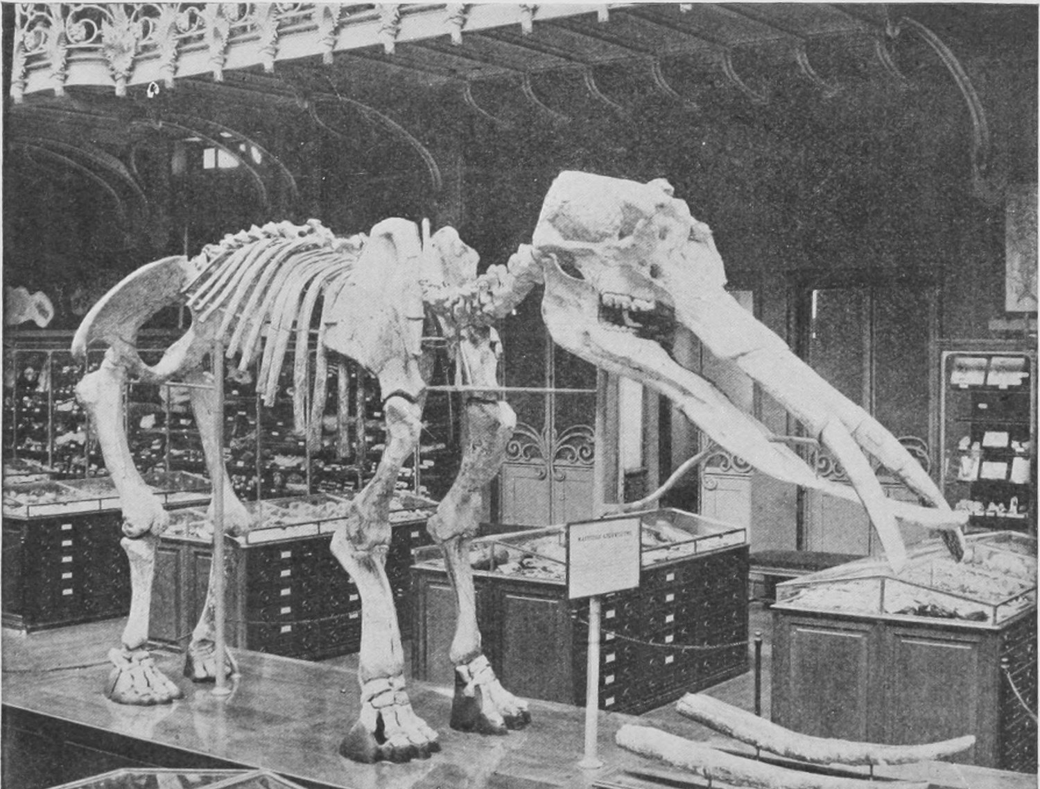
A further point in which the American mastodon is more like the ordinary run of mammals than are the elephants, is that it has front teeth —a single pair—in its lower jaw when it is quite young. These drop out in the American mastodons, but we have here a photograph (Fig. 79) of the skeleton of a much older mastodon, the remains of which were dug up in strata of the Middle Miocene (not only below Pleistocene, but below Pliocene and below Upper Miocene) in France. This skeleton is preserved in the Museum of Paris, where the photograph was taken. You will see that its head differs in many ways from that of elephants and the late American mastodon. It has an extraordinarily long lower jaw, with two tusks in it. A drawing of the side view of the skull is shown in Fig. 80, and you can see how the two horizontal lower teeth must have played between the two curious downwardly-bent tusks of the upper jaw.
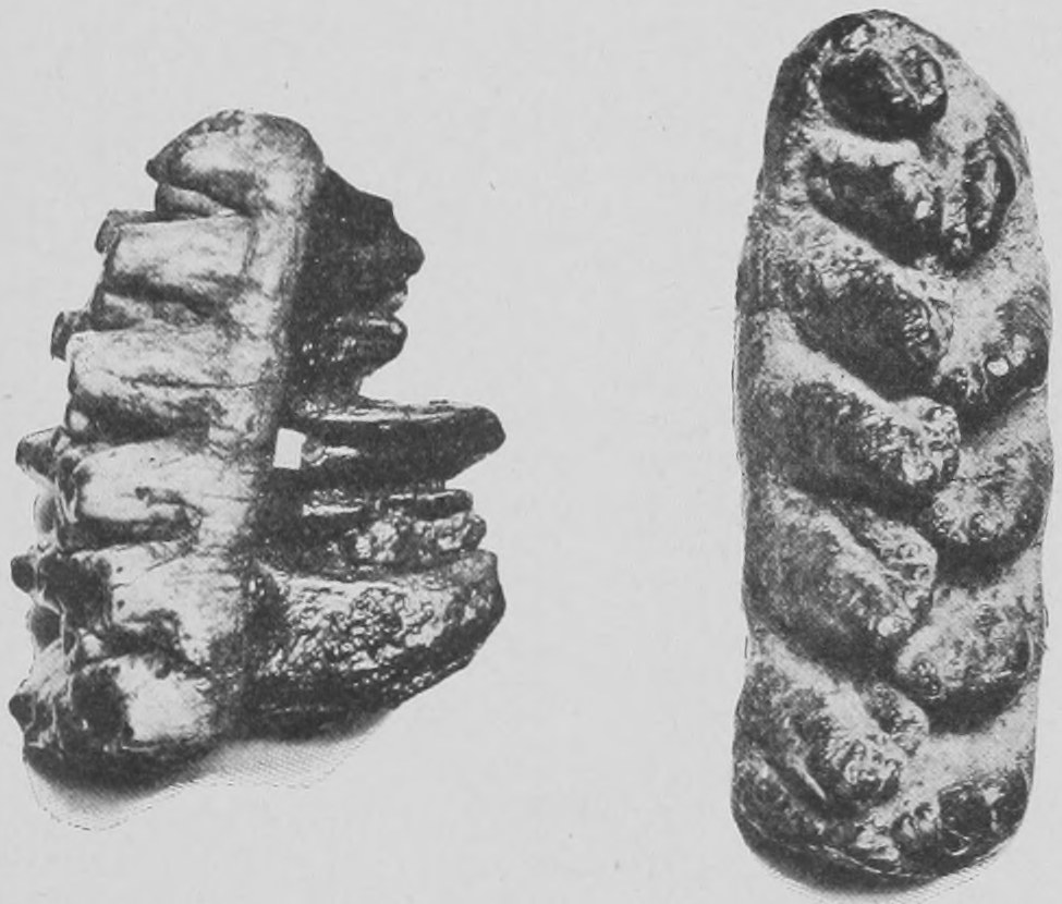
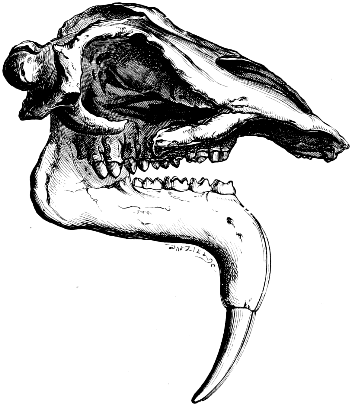
By way of parenthesis I must here mention a mastodon-like creature of the same age—which had no tusks in the upper jaw, but two huge tusks in the lower jaw, which is bent downward. This is the Dinotherium, found in the Miocene in Germany and other localities. It seems to have left no modern representatives, and is a sort of extinct side-branch of the elephant family. The big tusks of the lower jaw were probably used for raking up roots in the mud of rivers and lakes.
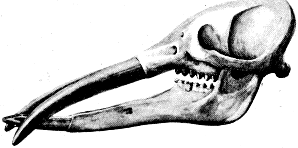
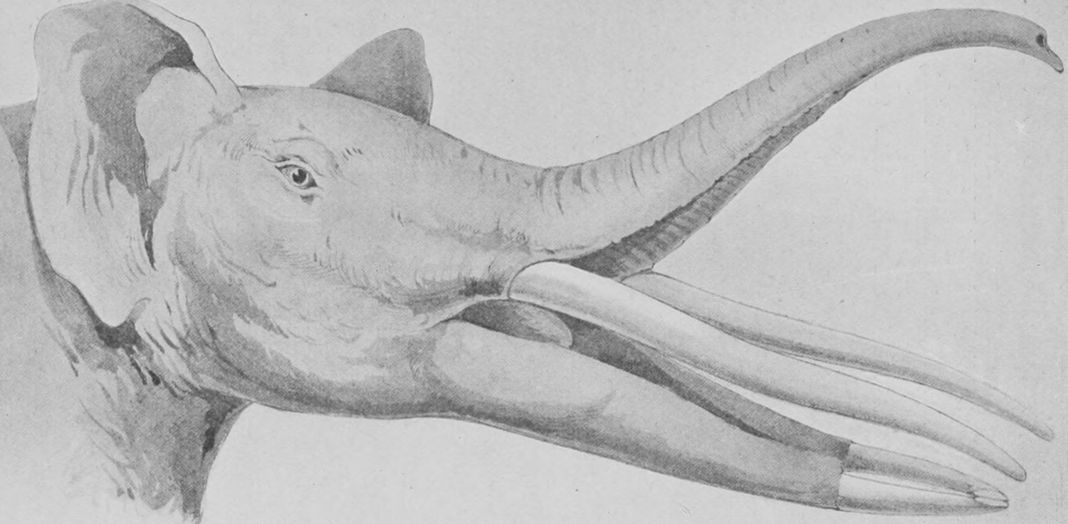
The Miocene mastodon, with the long lower jaw, is known as Tetrabelodon angustidens. The examination of its skeleton some years ago led me to the conclusion (as, indeed, was inevitable) that it could not have had a depending trunk like an elephant has and such as the short-jawed mastodons certainly must have had. Its "trunk" must have rested horizontally on the long lower jaw between the upper tusks—and was in fact not a "trunk" at all, but an elongated upper lip (Fig. 82)—representing the middle part of the upper jaw in a soft, flexible condition. It seemed to me probable that the elephant's trunk had originated in this way: namely by the great elongation, in the first place, of the lower jaw and upper lip and jaw, and by the subsequent shrinking of the lower jaw and bull-dogging of the bones of the face. Thus the elongated mid-part of the face—no longer supported by a long lower jaw—would gradually drop as the lower jaw grew shorter and shorter in successive ages, and at last it would hang down as a perpendicular trunk.
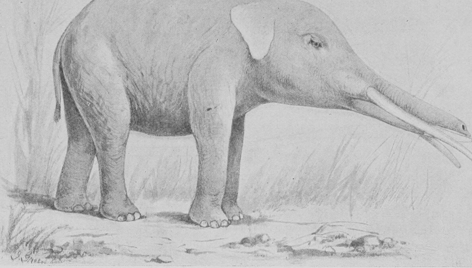
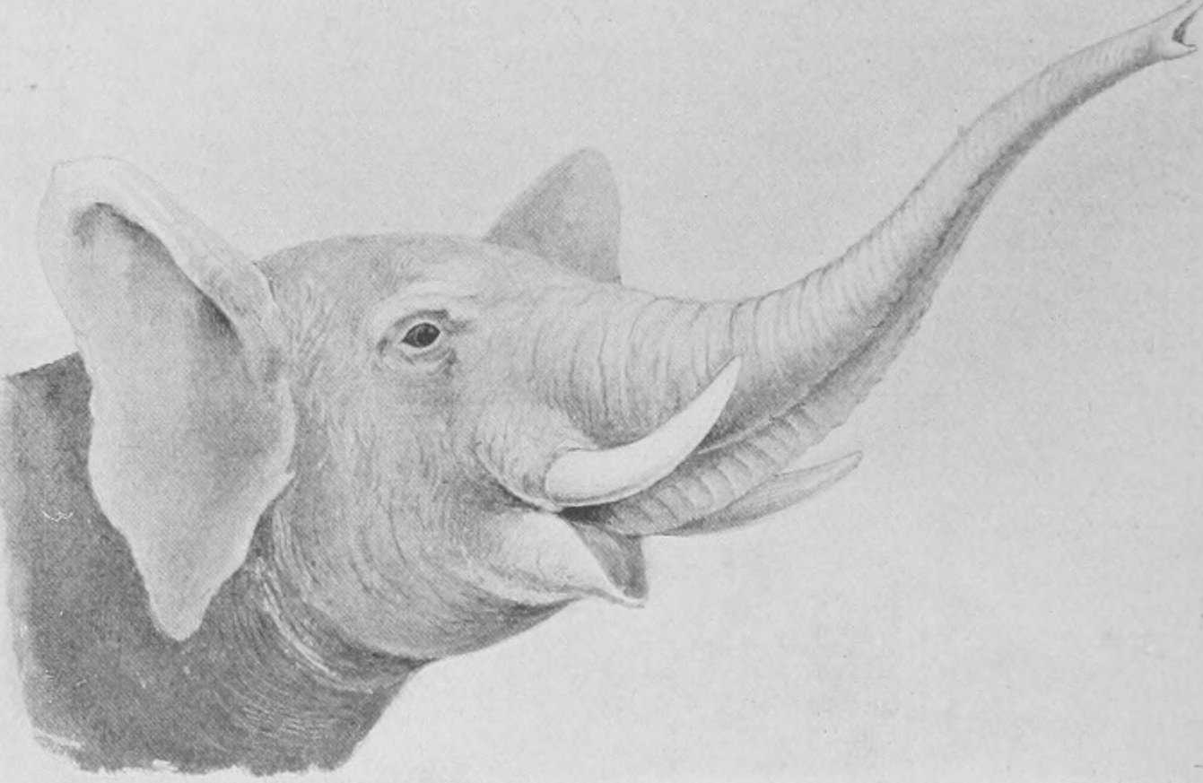
In Fig. 83 I have endeavoured to represent this long-jawed mastodon (Tetrabelodon) opening his mouth and rearing his flexible, boneless upper jaw as does the living elephant rear his trunk (Fig. 84). It is very difficult to form a definite idea as to how the Tetrabelodon made use of his tusks and horizontal "trunk." The upper tusks have a sharp edge along the inner face strengthened by enamel, so that it is probable that, working against the tough skin pads of the lower jaw, they would serve for cutting vegetable matter.
My friend Rudyard Kipling has given a different account of the origin of the elephant's trunk, which he declares was formed by the pulling of the nose of an unfortunate young elephant which, before the days of trunks, stopped to drink some water from a pool, and was seized by an enormous crocodile just about the nose.
The elephant pulled, and the crocodile held firm, and the result was the stretching of the elephant's nose till it became a trunk. This story was not told to Mr. Rudyard Kipling by the boy Mowgli of the Jungle Book, who I feel sure must have heard from the elephants the history as I have given it unless, as is not unlikely, they have forgotten all about the way in which their trunks grew, and would reject, as most men and women do, the notion that they have been derived by slow change in long ages of time from other and more simple animals.
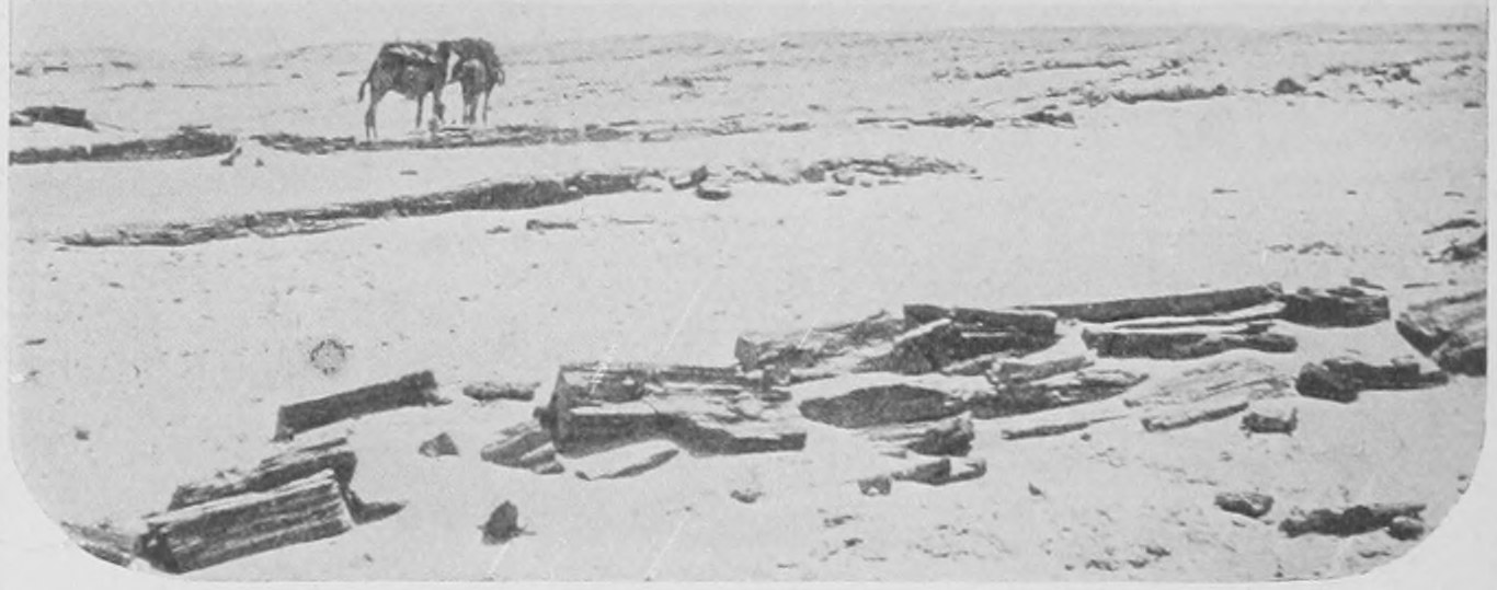
The history, suggested above, of the gradual production of the elephant in the later ages of the world's history from a long-jawed creature has been wonderfully confirmed by the discoveries made in Egypt within the past four years by my friend Dr. Andrews, who is one of the staff of the Natural History Museum. Dr. Andrews was in Egypt four years ago on account of his health and joined a party of the officers of the great survey of Egypt, organized by Lord Cromer, in a visit to the Great Western Desert, the rainless, sandy waste lying west of the Nile, not very far from what is now called the Fayum, and where in Roman days was the great Lake Meris—now dried up to a mere brine-pool, in the salt water of which the freshwater fishes of the Nile still live. The surveying party intended to determine the geological age of these sands, which stretch for hundreds of miles, often rising into cliffs which are cut sharp by the wind and show horizontal stratification. Some fragments of bone had been recorded from this region twenty years ago by the traveller Schweinfurth, and Dr. Andrews, who is a special expert and authority in the interpretation of fossil bones, was hopeful of securing some specimens for the Natural History Museum. He was rewarded far beyond his expectations. The party had to travel into an absolute desert waterless region, establishing a staff of camels which daily brought up water as far as three days' march into the sandy wilderness, returning with empty tanks on their backs to fetch more. In Fig. 85 is reproduced one of many photographs taken by Dr. Andrews. It shows the flat sandy desert with some fossilized lumps lying in the sand which are the remains of trees. The geologists determined that the sands in this region were of Upper Eocene and of Miocene age, and from them Dr. Andrews brought home some very interesting bones. These included remains of a more primitive mastodon than any as yet known and of an animal which he called Meritherium (after Lake Meris)—which is the connecting link between elephants and the central typidentate mammals. But the collection included also remains of great carnivores, of Hyrax of great size (like the Syrian coney), of Sea-cows (Sirenians), and of Tortoises, and a Snake sixty feet long. The Egyptian Survey has since in the most enthusiastic way sent further expeditions into this desert to collect the bones of the extinct animals half-buried there, and Dr. Andrews, by the direction of the Trustees of the British Museum and further assisted by a generous donation from Mr. de Winton, has twice again in succeeding years camped out in the desert and excavated the sands by the aid of a troop of native diggers.
In regard to the history of elephants, the upshot of Dr. Andrews' most important discoveries is that we find living here in the Upper Eocene period (older than the German or French Miocene with its Tetrabelodon) an elephant ancestor of the mastodon kind to which Dr. Andrews has given the name Palæomastodon. The skulls and many limb-bones of this interesting creature have been obtained, and are now reposing, some in Cromwell Road and some far away in the fine Museum of the Egyptian Survey in Cairo. In Fig. 86 a drawing (No. 4) is given of the skull of this Palæomastodon. The figure includes several other elephant forms. We have the skull and lower jaw of Tetrabelodon (No. 3), of the American mastodon (No. 2), and of the Indian elephant (No. 1). It will be seen at once how completely the Palæomastodon skull fills in the series leading back from the bull-dog-faced elephants with short jaws to ordinary mammals. It has a fairly long skull and long bony face, with two large—but not very large—downwardly directed tusks. The jaws are long, but the lower one not so excessively long as that of Tetrabelodon (No. 3), and the cheek-teeth are there in nearly full number—as many as five in each half of each jaw. These are well seen in the view of the lower jaw given in Fig. 87 (No. 2), where the condition of the lower jaw of Palæomastodon is clearly contrasted with that of Tetrabelodon (Mastodon angustidens, No. 3).
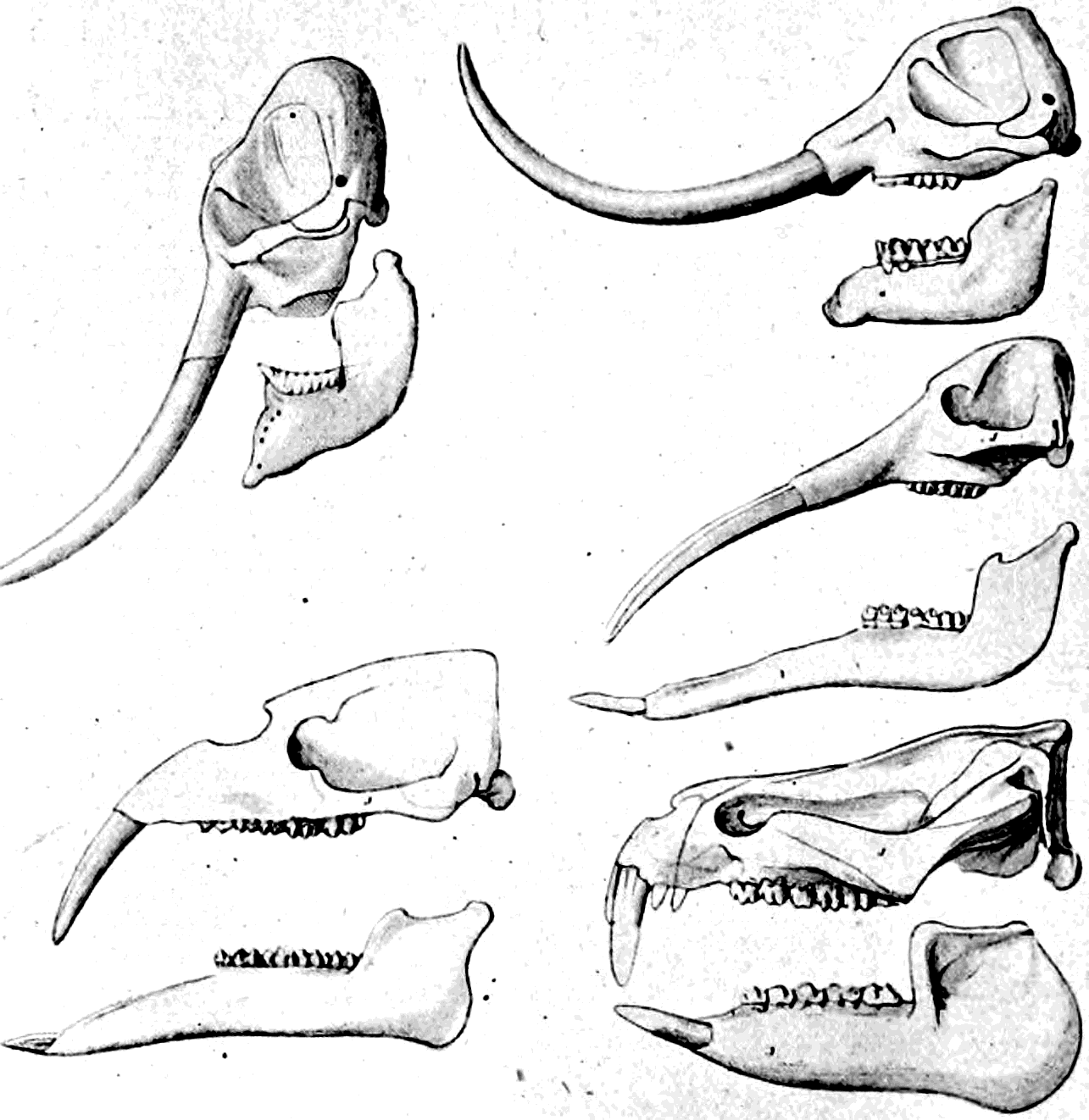
In Palæomastodon we have arrived, by passing as far back as the Eocene strata, at an ancestral elephant-like creature which serves to join the elephant stock on to more ordinary "normal" mammals. I should say that this beast was not so very big—about as large as a fair-sized horse. Dr. Andrews' great triumph, however, is the discovery of a somewhat smaller animal in the same deposits, which is undoubtedly an elephant, and yet at first sight has no resemblance to one and probably had no trunk at all, as certainly it had only small tooth-like tusks, unworthy of comparison with the great ivory columns of later elephants. This is the Meritherium seen in Fig 86 (No. 5), and more fully exhibited in Figs. 88 and 89. As is obvious at once, the skull of Meritherium does not suffer from "bull-dogging" at all; there is a fine, well-developed facial region, and the teeth are neither deficient in number nor greatly exaggerated individually. The "dentition" (that is to say, the enumeration of the complete series of teeth) approaches closely to that of the central mammals with typical dentition. In the upper jaw (as shown in Fig. 88) there are six front teeth or incisors, and it is the second of these on each side which is enlarged and is (so to speak) going to become the great tusk of the elephants. In the lower jaw there are four front teeth (see Fig. 87, No. 1). In the upper jaw we also find a small canine or dog-tooth; next the incisors and the cheek-teeth in both upper and lower jaw are fully represented, namely six on each side in each jaw—only one short of the type-number. And yet these cheek-teeth are quite obviously and recognizably mastodon teeth. They have the transverse ridges of the mastodon tooth (two or three) and are in other features like those of mastodons.
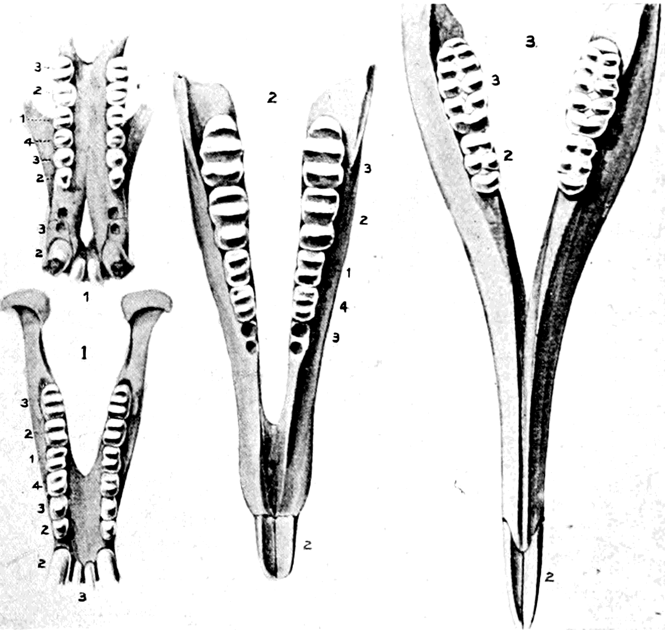
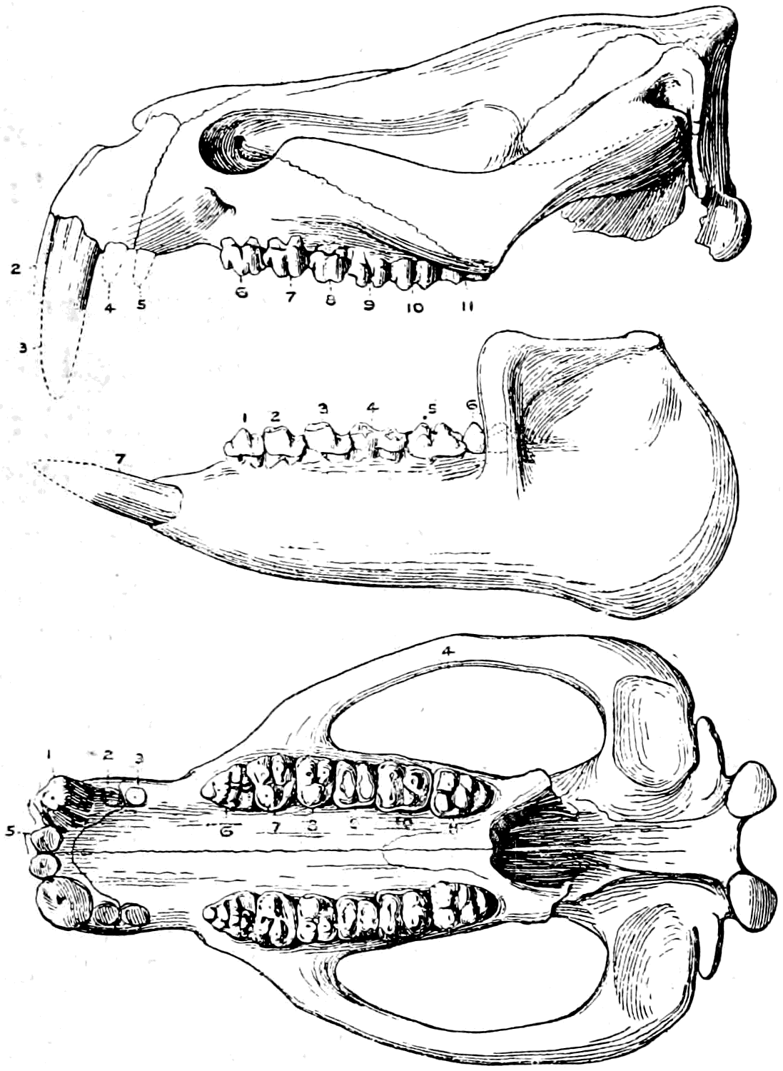
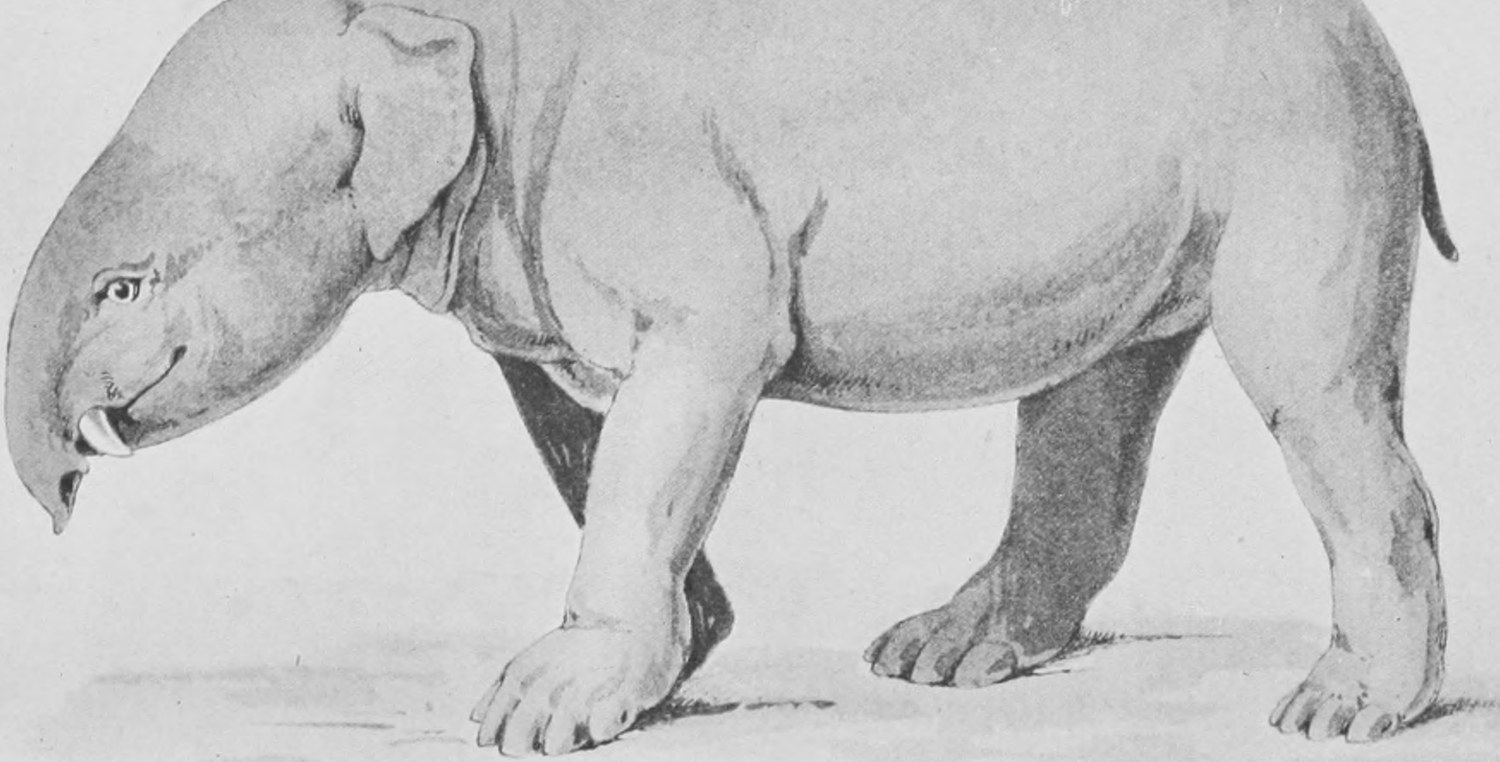
Here, then, we have arrived at a form which undoubtedly was closely related to the ancestors of all the elephants—if not itself actually that ancestor—and in it we see the origin of the elephant's peculiar structure. From this comparatively normal pig-like Meritherium, the wonderful elephant, with his upright face, his dependent trunk, and his huge spreading tusks, has been gradually, step by step, produced. And we have seen some, at least, of the intermediate steps—the elongation of the jaws and increase of the size of the incisors in Palæomastodon—carried still further in Tetrabelodon, and then followed by a shrinkage of the lower jaw and final evolution of the middle part of the face and upper jaw as the drooping, wonderful, prehensile trunk.
So much for the "great sagacious elephant" and his extinct relatives. Let us now turn for a few minutes to the most beautiful and the most helpful to man of all animals—the horse, nobler as he is bigger and stronger and more beautifully shaped, than man's other animal companion, the dog. The horse is curiously different from the central typical mammals in that he has only one toe on each foot instead of five, and further, in the complex pattern of his teeth. But immense numbers of extinct horses and horse-like creatures have been dug up, and we now know quite clearly all the stages leading from living horses back to four-toed and ultimately to five-toed ancestors. First of all I will put before you a photograph of a very beautiful model of an English thoroughbred (Fig. 90). There are a set of these models, both of horses and cattle, in the Natural History Museum: each is carefully modelled to one-fourth the size of nature. They were executed by a Hungarian artist for the exhibition at Buda-Pesth some years ago, and we ought to have such a series made now in England of samples of all the best breeds. It is the only way of keeping a really complete and satisfactory record, and such models of known horses and cattle, made to-day, would be of immense interest and value in fifty years' time. But they are costly things to make, and can only be undertaken by the rich owners of race-horses and pedigree bulls.
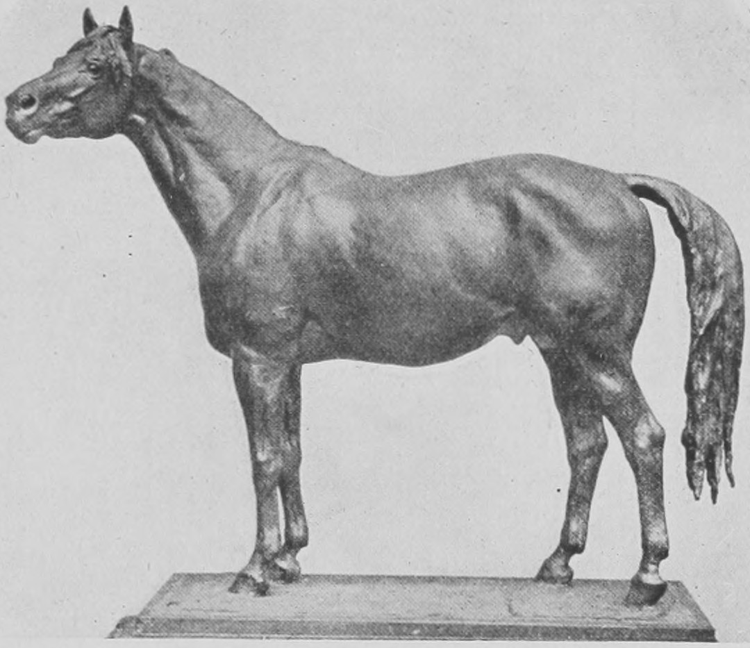
Fig. 91 shows us the fore and the hind foot of the horse. As is very usual with photographers and those who prepare drawings and lantern-slides, the artist has placed the hind-foot in front and the front-foot behind. The hind-foot (that on the left) shows the heel-bone or "hock" (the calcaneum) standing forth at the top of the ankle. Below you see the three bones which constitute, as in our toes and fingers, what is called the digit. Then there is a long bone, which is the meta-tarsal bone. In the front-foot the similar bone is called the meta-carpal. At the top of these are several short bones jointed together; these are the tarsus or ankle and the carpus or wrist (so-called "knee" of the horse's front-leg) respectively. You see, the horse walks on the very last joint of its toes, and keeps the foot and the hand upright, so that the heel is right above the toe instead of behind it, as in ourselves and the bears. On each side of the long bone of both fore and hind-foot you will see a small long bone, narrow and delicate. The nearer one of these delicate bones is not very clearly shown in the photograph, but still can be made out. These delicate "splint-bones," as they are called, are all that remain in the modern horse of two additional toes. There was a time when horses had three toes—far back in the Miocene strata we find horses which had three well-developed toes, each with a hoof resting on the ground (the Mesohippus and Anchitherium), and earlier than that we find a horse-like creature (Hyracotherium) with three nearly equal-sized toes on the hind-foot and four on the front foot (Fig. 92). In the Pliocene we find a three-toed horse in Europe known as the Hipparion (and a similar kind is dug up in America), which had three toes on each foot; but the side toes were getting small, were in fact like the "petti-toes" of the pig, and of cattle, and of the reindeer. They did not touch the ground (Fig. 93), and were evidently on the way to disappearing, leading to the single-toed modern horse, with its splint-bones, as the sole representatives of the two outer toes. Occasionally living horses are born with two complete little toes provided with hoofs and attached to the splint-bones, one on each side of the big central toe, "throwing back," as the term is, to their three-toed ancestors. Beyond the stage, with four equal toes on the front foot and three on the hind-foot, which is exhibited by a quite small horse-like creature—the Hyracotherium shown in Figs. 94 and 95—we can trace the pedigree of the horse to a five-toed ancestor, the Phenacodus (Fig. 96). The later stages of this history, from the Mesohippus to the modern horse, have been traced by very abundant fossil remains of many steps or stages in the gradual change. Not only has there been a gradual change from the three-toed to the one-toed condition, but there has been a great increase in size, and moreover the cheek-teeth have gradually become more and more complex in the pattern which they show when worn down. In Fig. 97 crowns of the cheek-teeth of the Mesohippus are represented, and in Fig. 98 the crown of an upper molar of a recent horse. There are a great number of interesting details in the history of the changes of the teeth and toes of the ancestral series of horses which it is not within my scope to describe here, but they may be studied on specimens of a variety of ancestral horses which have been set out for the purpose in the Natural History Museum.
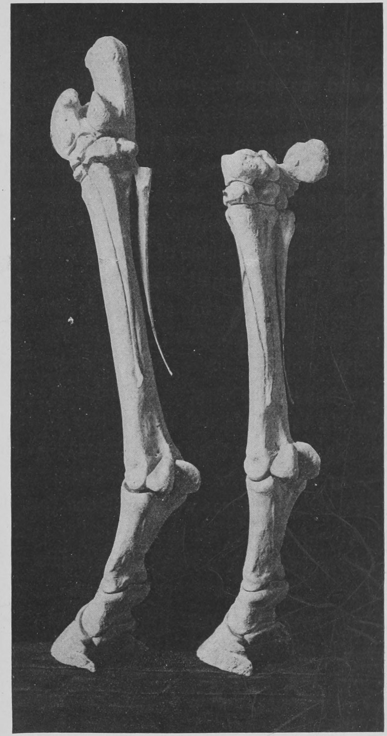
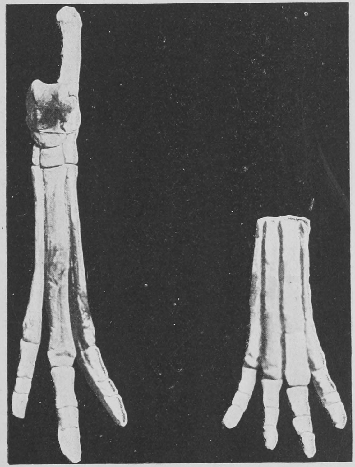
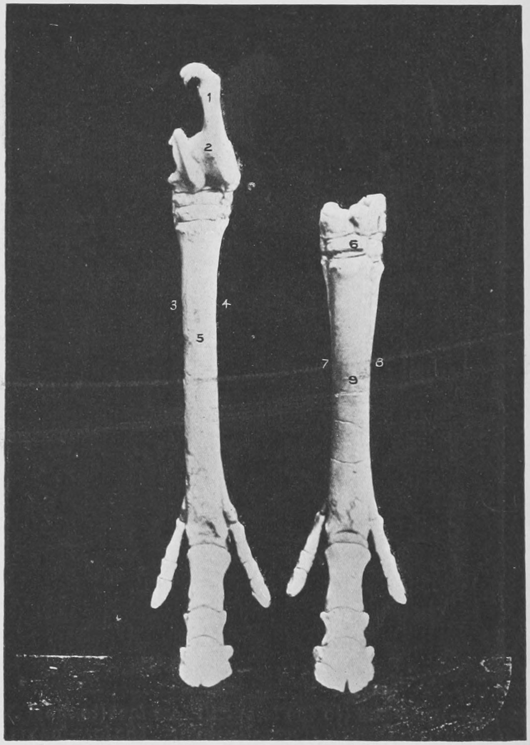
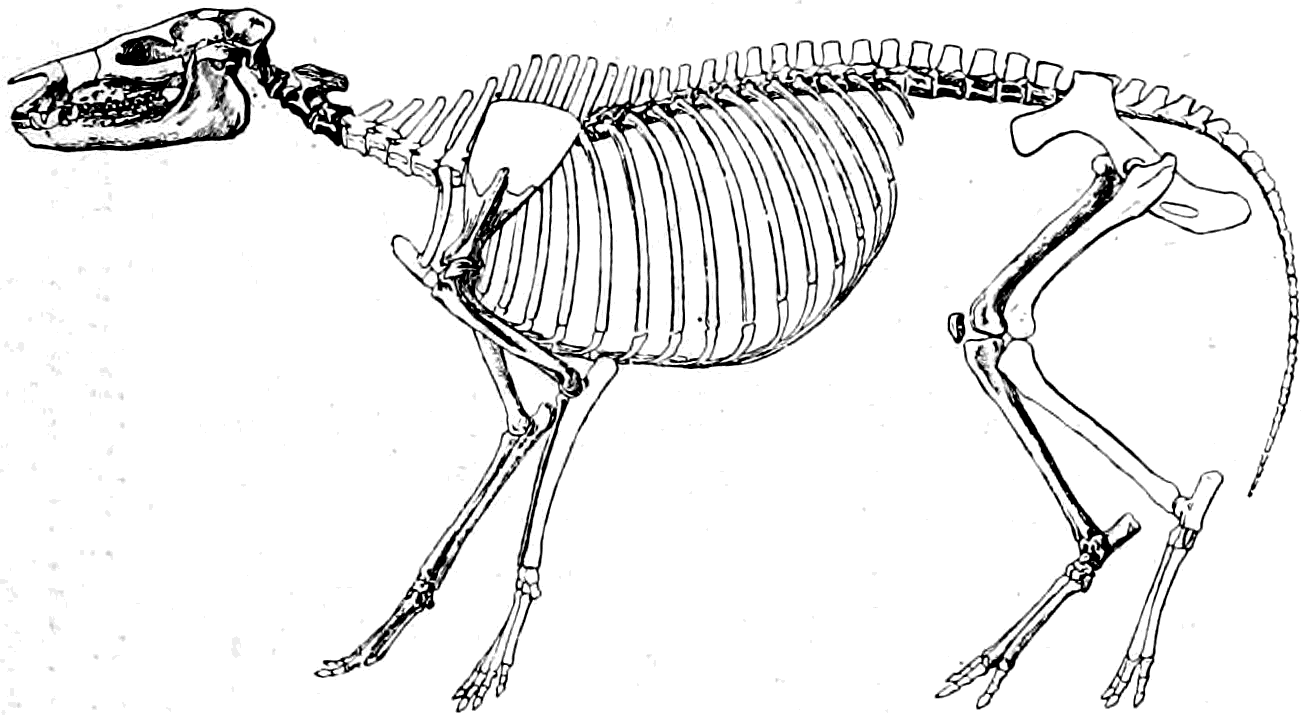
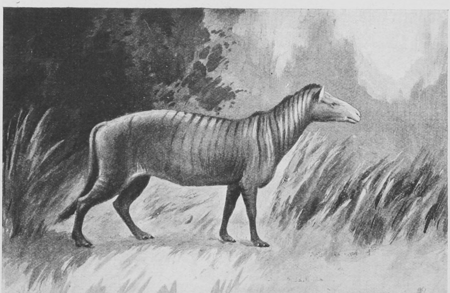
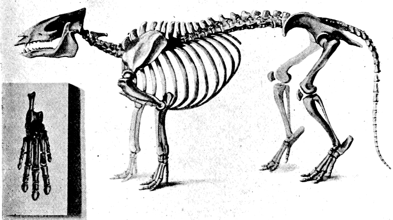
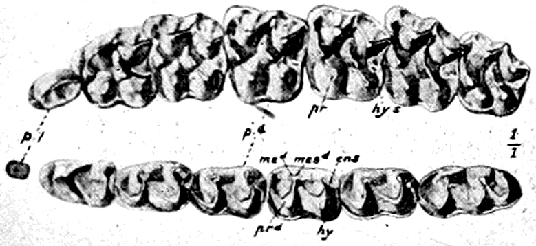
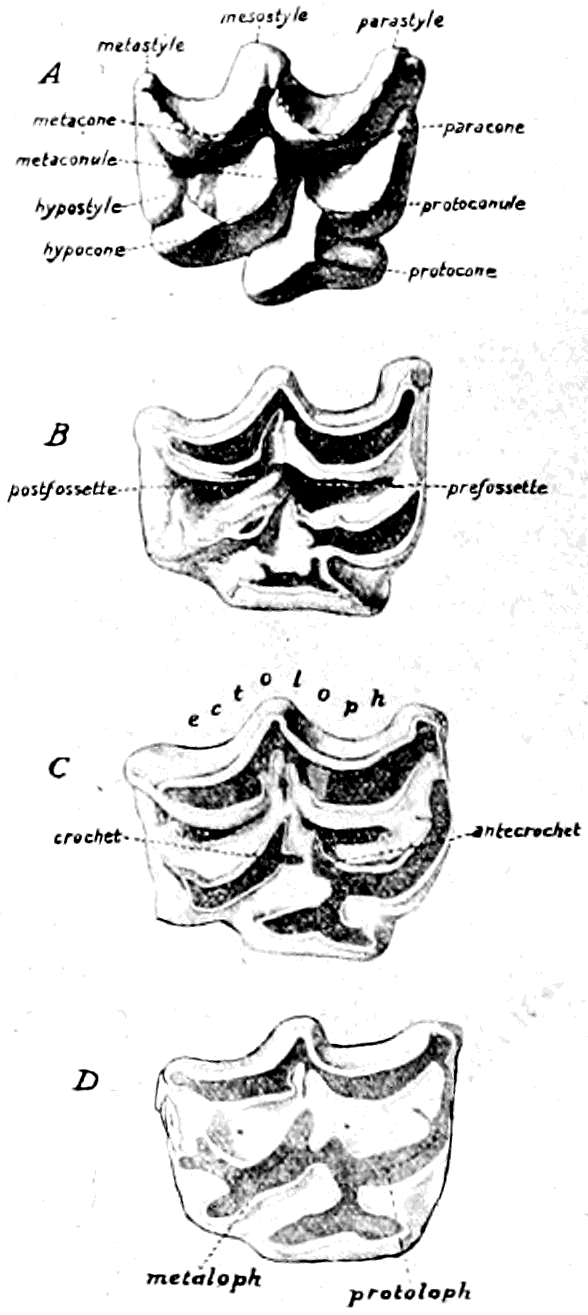
The rhinoceroses of to-day—the unicorn or Indian rhinoceros and the two-horned African rhinoceroses, one with a pointed upper lip and the other with a square, broad mouth—have been preceded by a whole regiment of extinct rhinoceroses, whose bones and skulls are dug up in the Pleistocene, Pliocene and Miocene strata. In Fig. 99 is represented the complete skeleton of the commonest kind of fossil rhinoceros, the skull of which was dug up in London the other day and is shown in Fig. 5. This rhinoceros had a hairy coat like the mammoth, and is found sometimes with the mammoth in frozen gravel in Siberia. The living rhinoceros most like it is the African square-mouthed rhinoceros or Burchell's rhinoceros (Rhinoceros simus), misleadingly called sometimes the white rhinoceros (Fig. 100). Many of the extinct kinds of rhinoceros had two horns, one behind the other like the African rhinoceros. The horn of the rhinoceros is truly horny in substance, and fibrous. It is not bone, as are the horns of stags, nor has it a core of bone as have the horns of sheep, cattle and antelopes. Sometimes, however, there is a fairly big boss of bone, which forms a sort of base or pedestal for the horny horn. One great extinct beast (the Elasmotherium), allied to the rhinoceroses, had a great horn, carried on a huge boss on the middle of its head instead of on the nose, whilst in the Miocene of North America complete skeletons have been found of an enormous creature allied to the rhinoceroses, but having a pair of horns, perched side by side on the nose, instead of one in the mid-line, or two placed one behind the other. The skeleton of this great beast, called Titanotherium, is shown in Fig. 101, and in Figs. 102 and 103 photographic views of the skull are given.
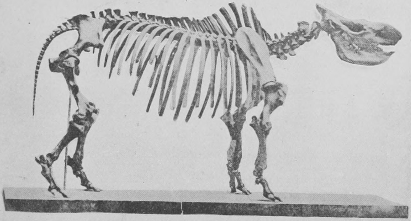
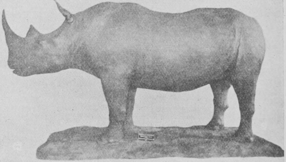
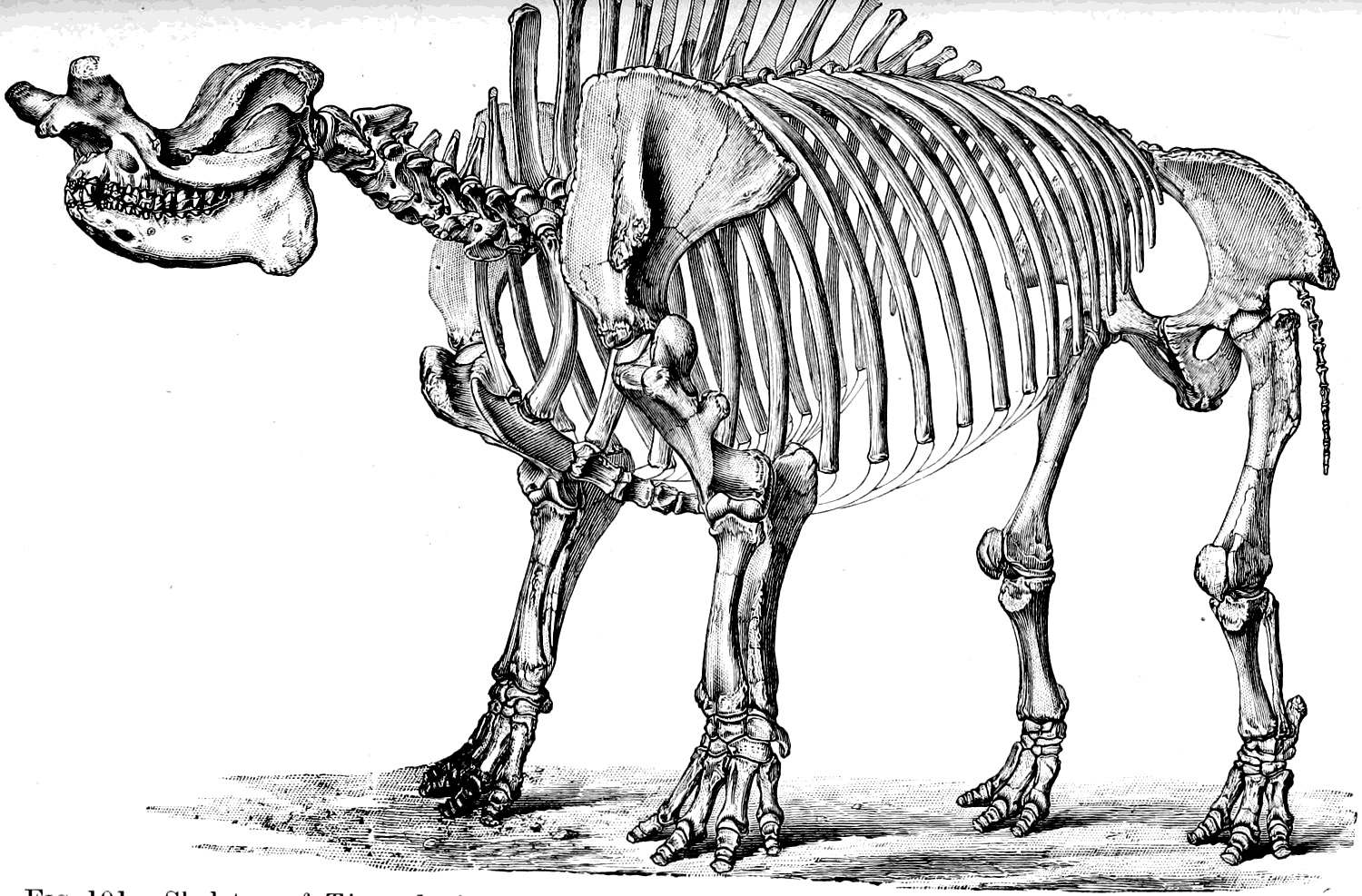
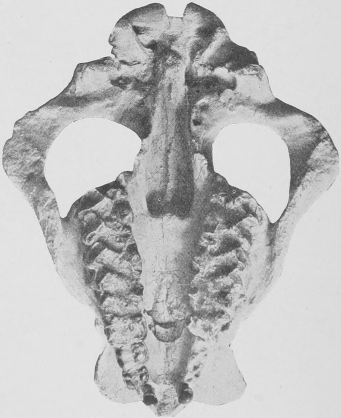
As large as the rhinoceros, but having a very different arrangement of the bones of its wrists and ankles, and very different teeth and horns, are the extraordinary creatures known as Dinoceras, whole skeletons of which have been disinterred from the Upper Eocene of Wyoming in the United States. As many as two hundred individuals were studied by Professor Marsh, who has written a large treatise on them. These creatures had three pairs of horns on the top of the head (Fig. 104) and a pair of great tusks formed by the enlargement of the upper canine teeth. The horns are outgrowths of the bone of the skull and were probably covered by hardened skin. The probable appearance of this creature in life is shown in Fig. 105.
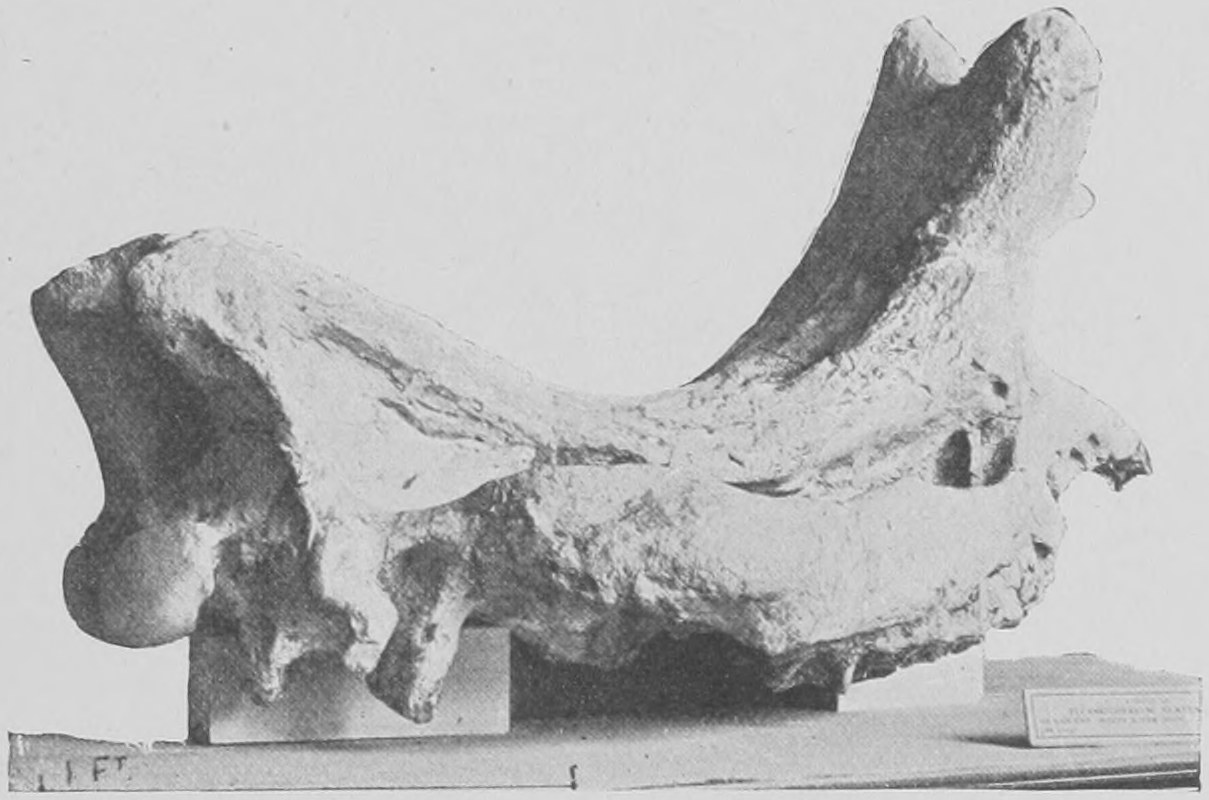
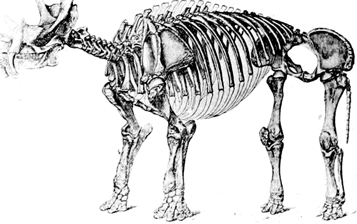
A very interesting fact has been observed about the brains of these most ancient big mammals, viz., the Dinoceras occurring so far back as in the Upper Eocene, and the Titanotherium of the Lower Miocene. We can get castings from the interior of the skulls and compare them with those of recent rhinoceros, hippopotamus and horse (Fig. 106), and it is found that although Dinoceras and Titanotherium were bigger than the largest rhinoceros of to-day, yet they had quite small brains, not more than an eighth the volume of that of the recent big animals. The subject has not been so fully looked into yet as it deserves, but it seems that modern animals, the animals which have survived, have much bigger brains than those which died out in the Eocene and Miocene times, and it is probable that they have survived to a large extent because of the value to them, in the struggle for existence, of the bigger brain.
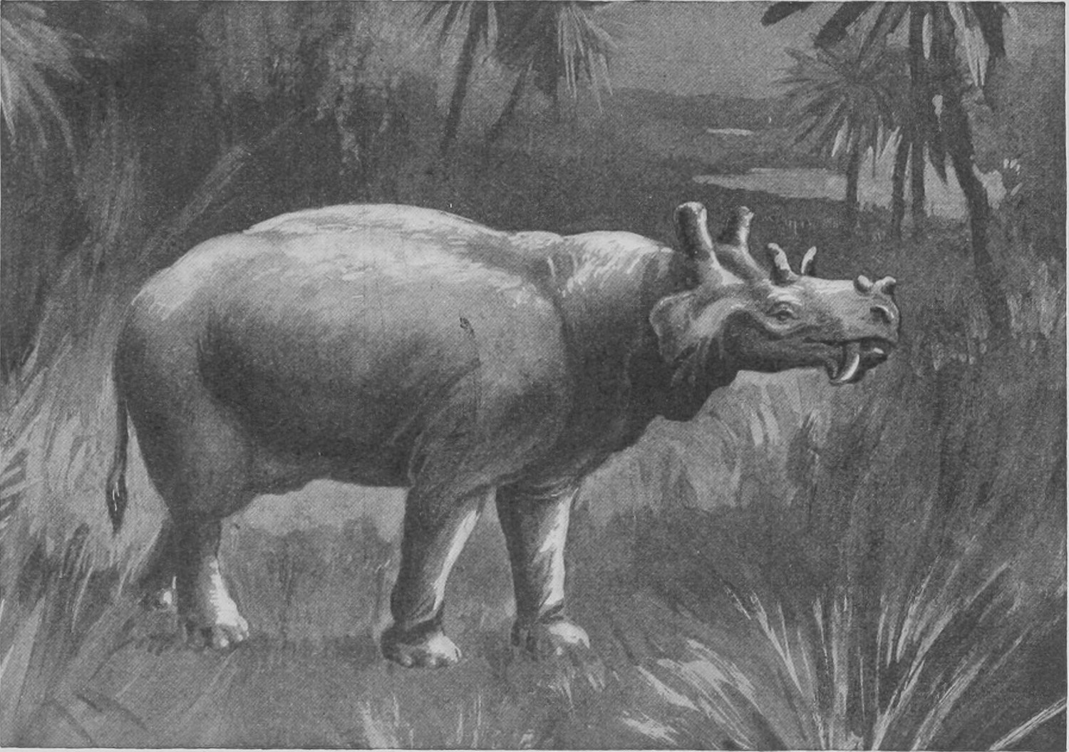
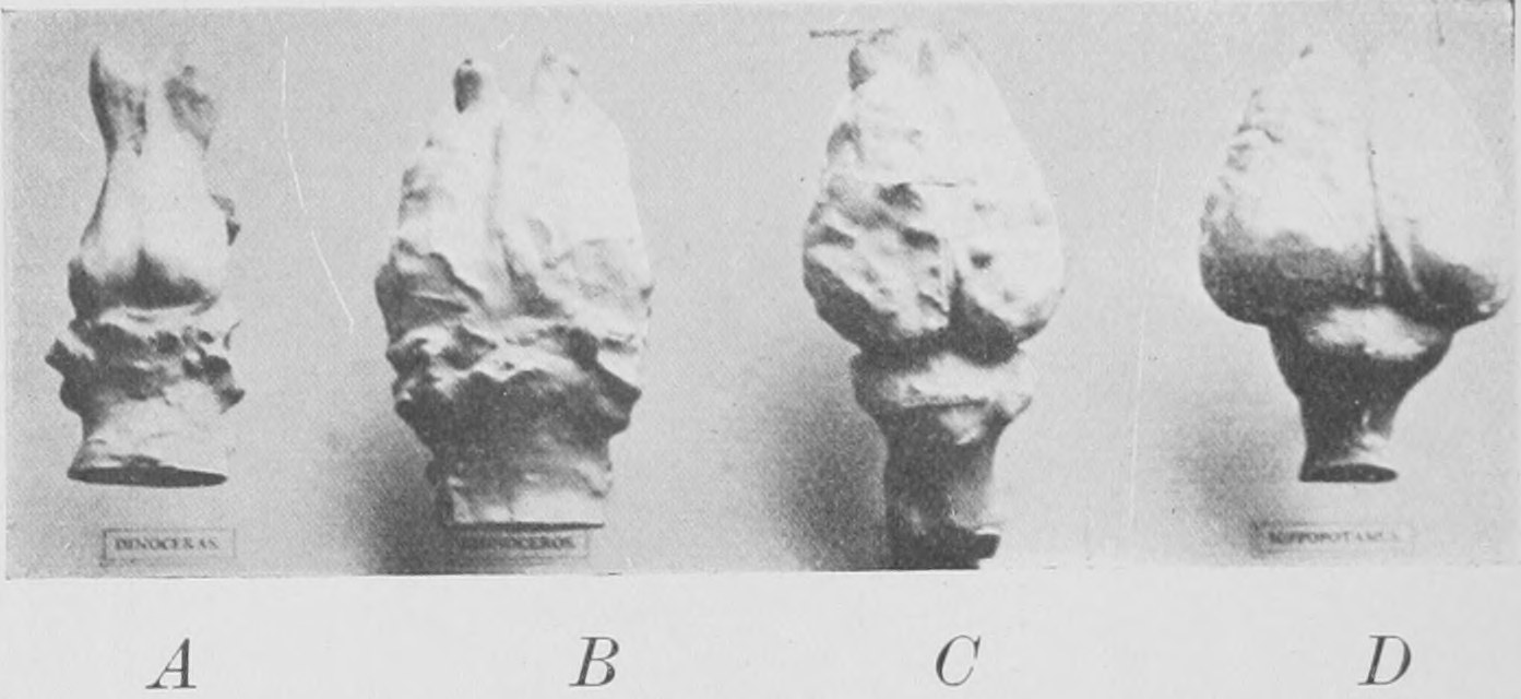
It seems that a small brain may serve very well to guide the great animal machine in established ways, but in order to learn new things in its own lifetime an animal must have a big brain—indeed, a very big brain. And the kind of animal which can learn—that is to say, can be educated—will, in the long run, beat the kind which has too small a brain to be capable of learning. This is the significance, not only of the big brains of recent rhinoceros and horse as compared with those of Titanotherium and Dinoceras, but it is also the significance of the big brain of man, which is far bigger than that of any other animal in proportion to the bulk of his body and limbs.
Another huge horned animal has quite lately become known which in some ways resembles Titanotherium and Dinoceras, but has to be kept apart from them on account of being really unlike them in its teeth and skull and feet-bones, although having a general resemblance to them in outline and bulk. This creature was found only three years ago in the same Upper Eocene sands of the Egyptian Fayum from which Dr. Andrews obtained the ancestors of elephants. The skull of this most strange animal is shown in Fig. 107, and a representation of what we suppose it looked like in life is given in Fig. 108. This wonderful beast was discovered by Mr. Beadnell of the Egyptian Geological Survey, and the name Arsinöitherium was given to it by him because the Egyptian queens of Greek race—named Arsinöe—had a palace near where the bones were dug up. Two thousand years ago many parts which are now sandy desert were well-watered and under cultivation. The drawing given in Fig. 107 is prepared from a skull in the Natural History Museum, where we have brought together portions of several other skulls and the complete set of bones of the skeleton dug up, some by Dr. Andrews and some by the energetic officers of the Egyptian Survey. The huge pair of horns are entirely bony outgrowths of the nasal bones, and are hollow. A small second pair of horns lies behind them. Probably in life the big horns were clothed with a horny case like the horn of a bull or antelope. The teeth are most remarkable, since they form a complete series, without a break, and are present to the full number—seven cheek-teeth, a canine and three incisors on each side in both upper and lower jaw—wonderfully graduated in form and size.
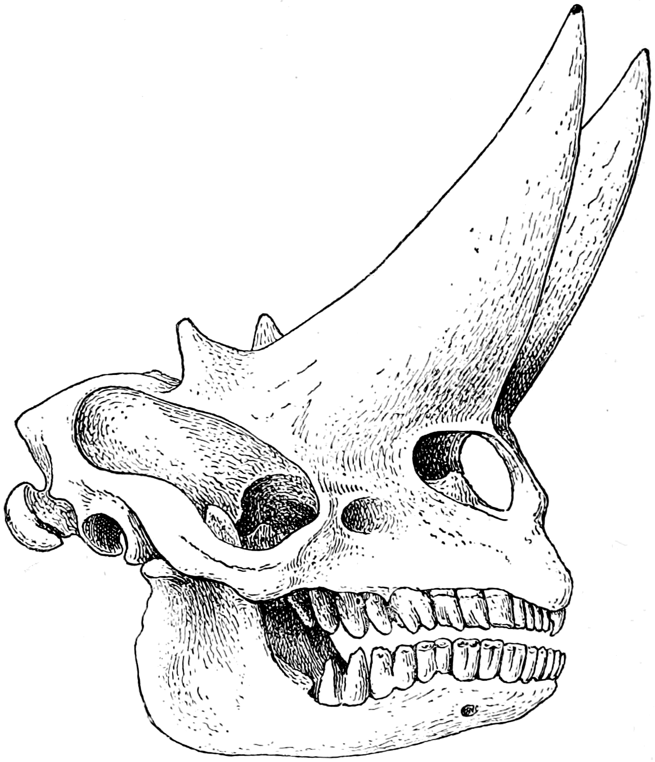
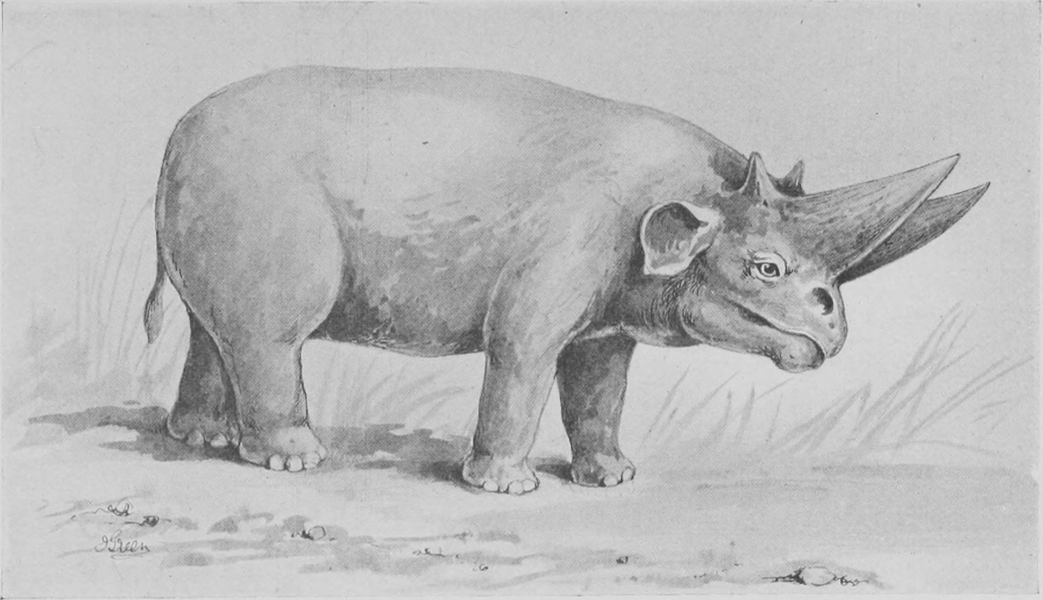
A complete account and illustrations of the remains of this most remarkable beast, the skull of which alone is nearly three feet in length, will soon be given by Dr. Andrews in a large volume on the extinct animals obtained from the sands of the Egyptian Fayum which is now in preparation and will be published by the Trustees of the British Museum.
Chapter Four: Extinct Giraffes and the Okapi—The Giant Sloths of South America and the Giant Kangaroos of Australia
There are a vast number of mammalian extinct animals, related to the cattle, sheep, goats, antelopes, deer, lions, bears and hyenas of to-day, and other less-known warm-blooded hairy quadrupeds, besides many, such as the Dinoceras and Arsinöitherium, which have left no successors like themselves to represent them in our days. Of both kinds, those which have quite died out and disappeared and those which have representatives alive to-day, you may see the bones and skulls in the Natural History Museum. I have not space here to speak of more than a few extinct creatures, and will at once ask you to look at some members of the group which to-day is familiar to us through the beautiful giraffe of Africa—the camel-leopard, the spotted, long-necked creature which will very soon be killed out by the intrusion of civilized man into the African wilds.
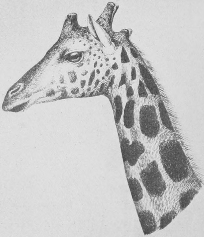
We have already seen a photograph of the giraffe in the first lecture, with its long neck reaching forward and forming a continuous line with the back. To-day I show you a sketch (Fig. 109) of what Sir Harry Johnston calls the five-horned giraffe. The ordinary giraffe has a pair of short bony outgrowths or so-called horns on the parietal region of the skull and a single horn of similar character between the eyes. But the five-horned giraffe has an additional short pair of outgrowths at the back of the head. All these "horns" in the giraffe are covered in life with living skin. There is no horny covering to them, nor do they grow through the skin and project as naked bone, as do the antlers of deer. The skull of the five-horned giraffe is seen in Fig. 110. Whilst the two hinder knobs, or horns, are real "outgrowths" of the skull, the chief horns (the median and the large pair, of which only the left-hand one is visible in the photograph) originate as separate bony pieces, which, after growing for a time as distinct bones, join tightly to the skull. Sir Harry Johnston shot the five-horned giraffe in the great "reserve" or protected area formed by the crater of the extinct volcano Mount Elgon—some five miles across—in the British Central African Protectorate of Uganda. In less than three weeks from the day on which he shot these specimens he was in London, and brought the skins and skulls of the specimens to the Natural History Museum! Central Africa, under the equator, can now be reached in that short space of time.
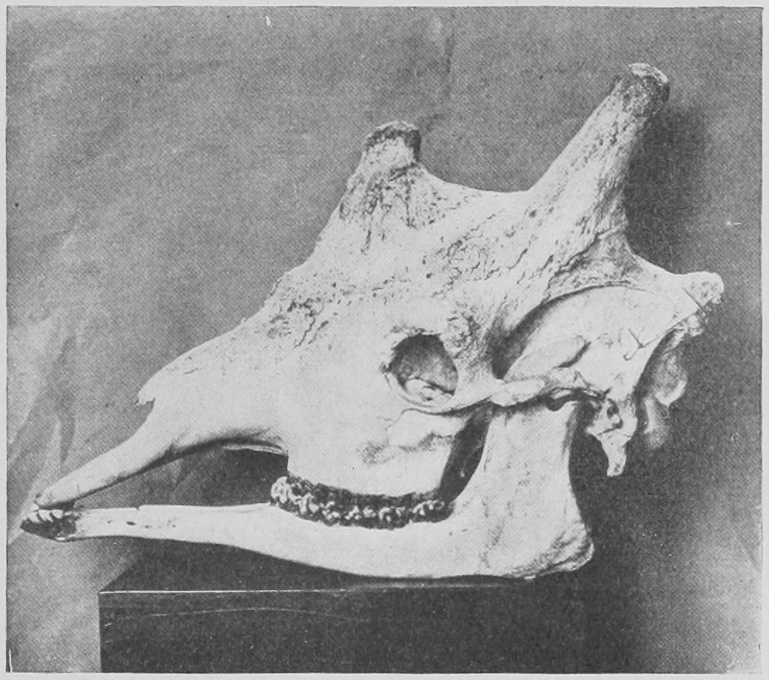
In Miocene times there were other large animals allied to the giraffe, but without so great a length of neck. The giraffe family have double hoofs, like the cattle, sheep, antelopes and deer, to which they are allied—not single or triple hoofs, like the horse family. Besides their peculiar and very primitive horns they have another small but definite peculiarity. The outermost of the group of eight front teeth in the lower jaw corresponds in position to the canine of the pig and other typi-dentate animals (animals with "typical" dentition, that is to say, little altered from the form and arrangement in early mammalian ancestors). In the cattle, sheep, antelopes and deer, this tooth has a quite simple chisel-like crown, like that of the incisors. But in the giraffe it is very peculiar: the crown is divided by a slit into two halves, each of which is large and broad. It is described as bi-foliate (see Fig. 111). No other mammalian animal was known with this peculiar shape of this particular tooth among living animals until the other day. But a great extinct animal from India, the Sivatherium (Fig. 112), with much larger horns than a giraffe, has this same bifoliate canine on each side in the lower jaw (Fig. 111), and is shown, by this and other facts in its structure, to be clearly related to the living giraffe. Another creature from the Miocene strata of the isle of Samos—the Samotherium (Fig. 113)—has also the bifid lower canines, and is closely allied to giraffes. The entire skeleton of a giraffe-like animal with a moderate length of neck has been found in Miocene beds in Greece, and is called the Helladotherium (Fig. 114). Naturalists were, therefore, deeply interested when Sir Harry Johnston obtained, some four years ago, from the borders of the Congo State where the great Congo forest approaches the river Semliki, which separates Congo-land from Uganda, a skin and two skulls of a new animal—the Okapi—which he rightly surmised to be a second living genus, or kind, allied to the giraffe. I gave the name Okapia to Sir Harry Johnston's new animal; it is stuffed and exhibited in the Natural History Museum. Like the giraffe, it has paired hoofs and a rather long neck, but it is striped on the legs and haunches, instead of being spotted. The most decisive point about its relationship is found in the canines of the lower jaw which, although small in size, are bifid or bi-foliate, as are those of the giraffe (see Fig. 111). Our specimen (Fig. 115) is about as big as a large stag; it has no horns, and is not adult. It is probably a female; the male, we now know, has a pair of horns (Fig. 116), and is extremely close, in the details of its skull, to the Samotherium (Fig. 113). Some fifteen specimens of this new and rare animal have been received in Europe since Sir Harry Johnston discovered it; it is probable that there are two species, a smaller and a larger, living both in the forests of the Congo in the centre of Africa. As they live in these immense dark gloomy and damp forests they are very difficult to shoot or to catch, and moreover they are not abundant. The natives cut the striped skin into girdles and bands for ornament. Two of these were sent home by Sir Harry Johnston before the animal was known, and were described as coming from a new species of zebra which was named Equus Johnstoni by Dr. Sclater (see Fig. 117).
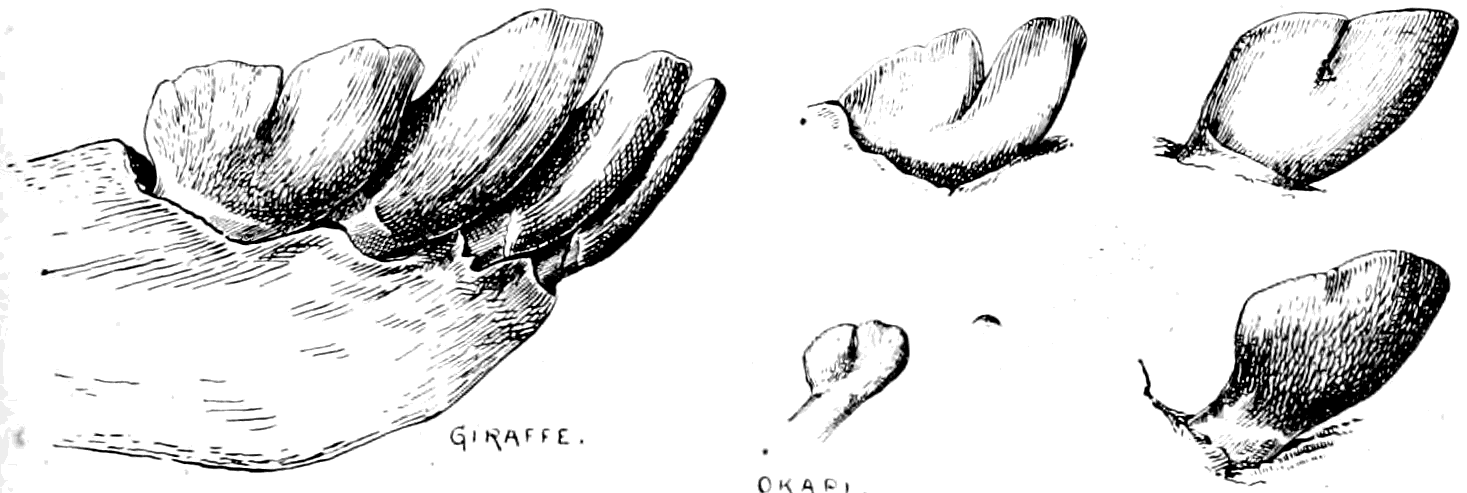
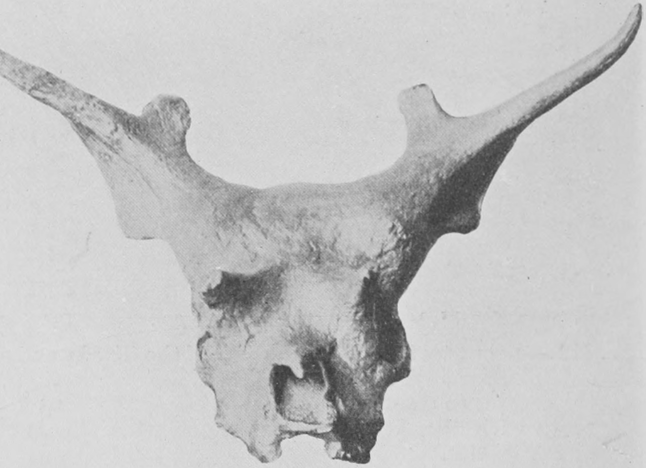
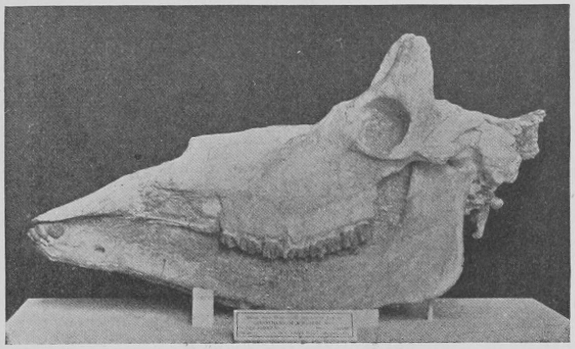
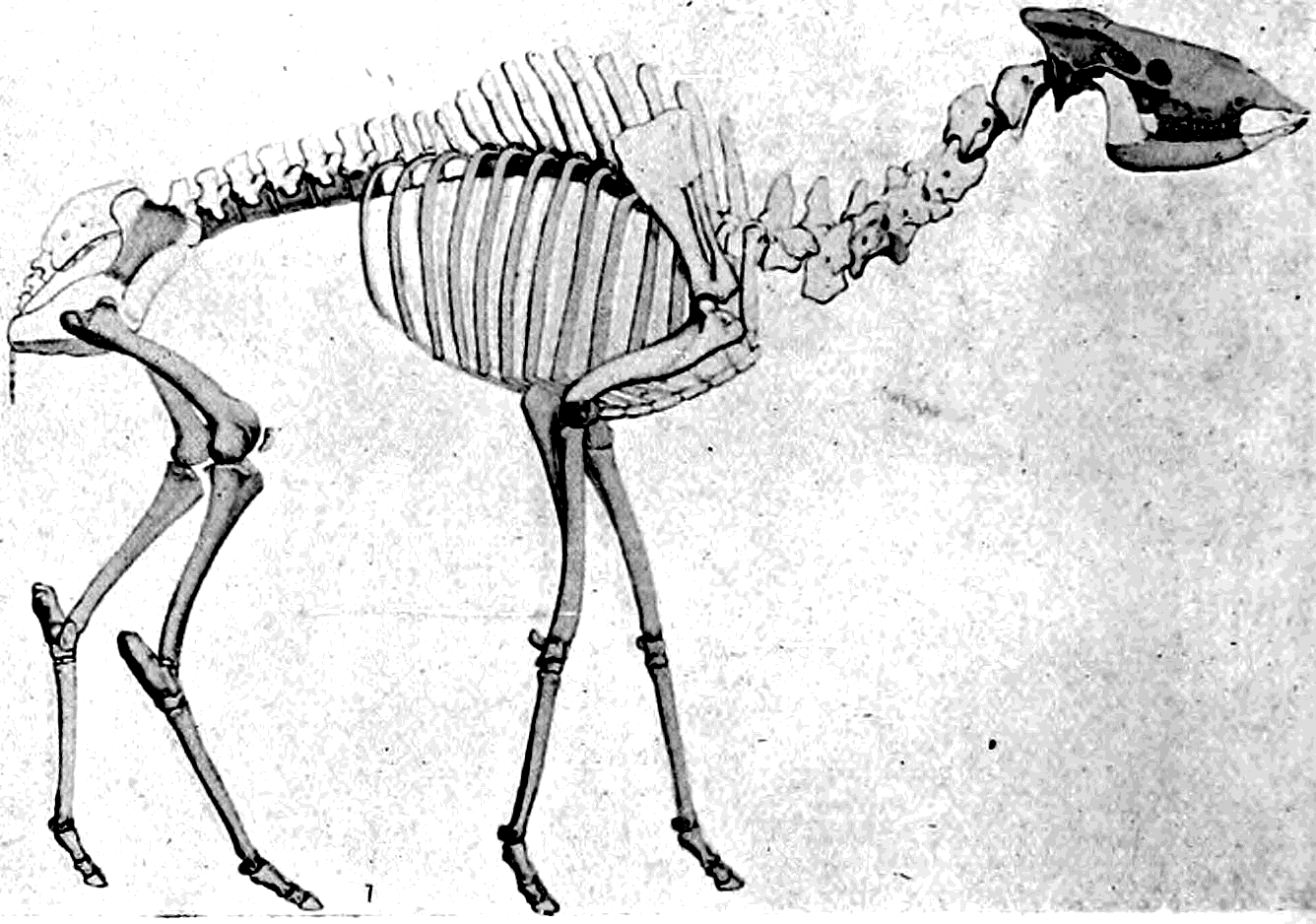
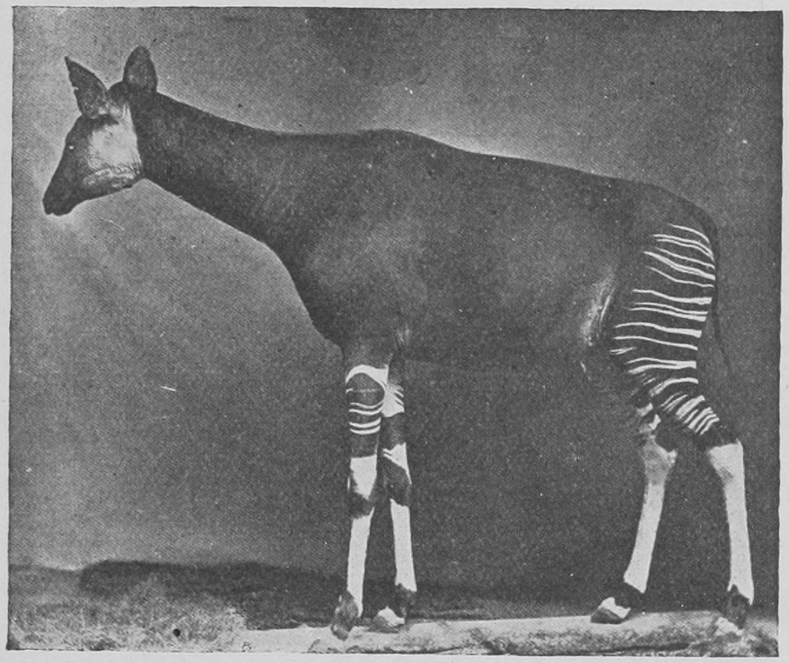
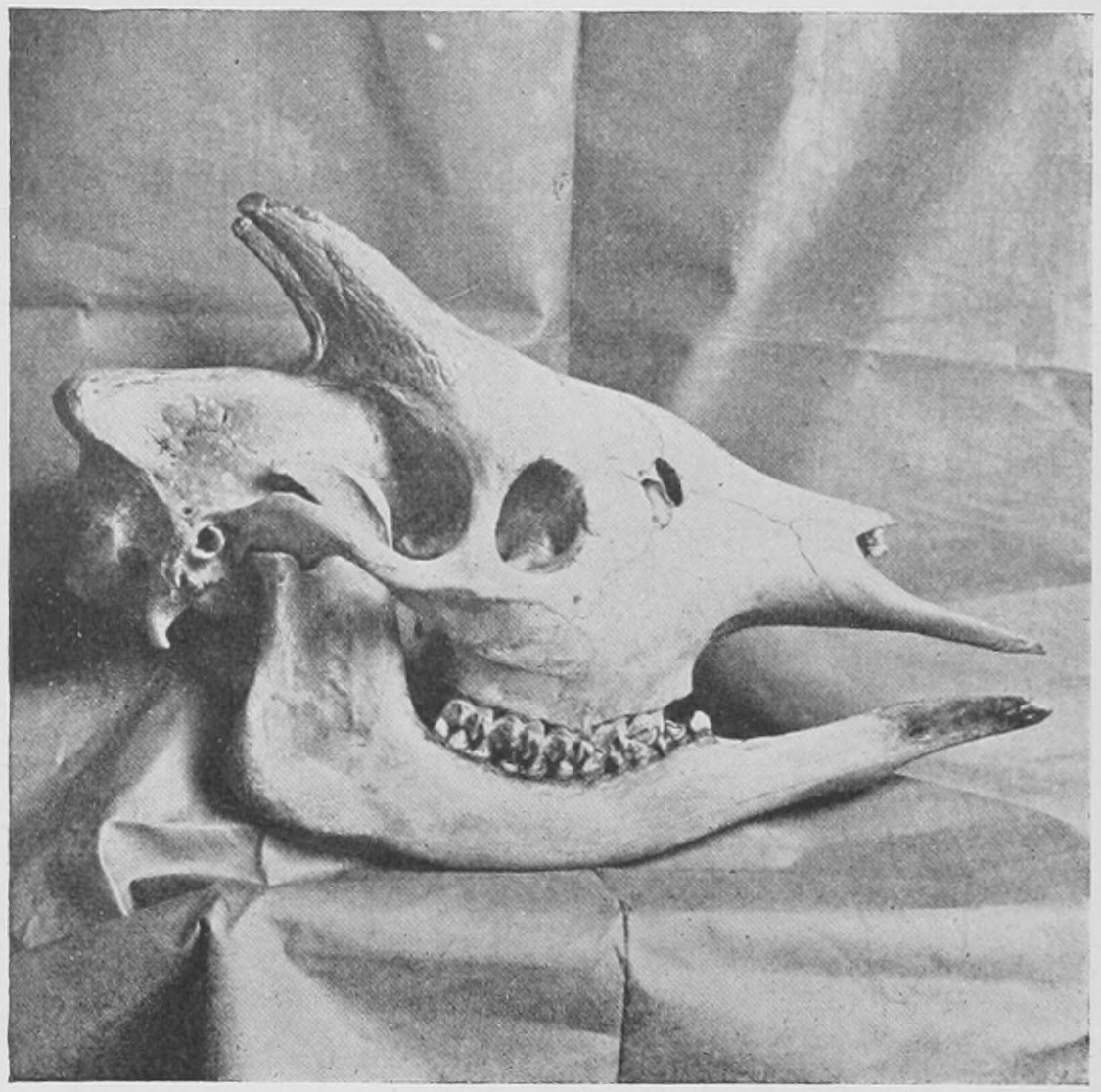
Some people, on account of the Okapi being striped somewhat like a zebra, whilst it has the double hoofs of giraffes and also paired horns, have supposed that it might be a hybrid or "mule" between a zebra and a giraffe. This is, however, a supposition which every naturalist knows to be quite out of the bounds of remotest probability. It is a fact that "mules" or hybrids never are produced by animals living in their natural conditions, except in a few rare cases among aquatic animals whose eggs are fertilized in the water after they have been laid. And no one has ever produced, even in captivity, a hybrid between any creatures so unlike each other as a double-hoofed and a single-hoofed mammal.
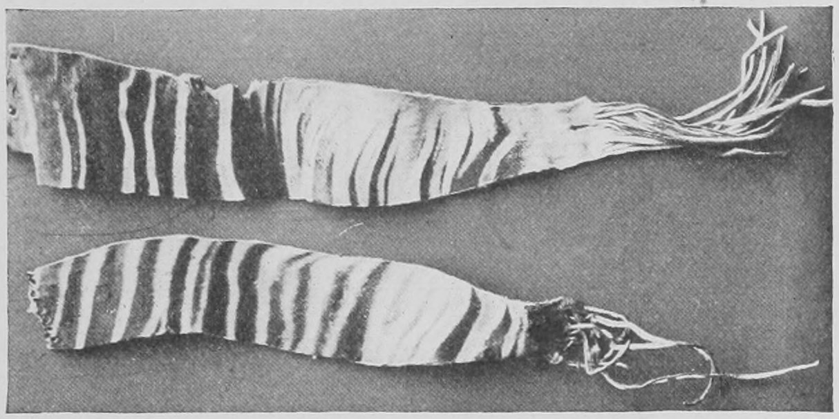
There are a good many instances in which small living animals were represented in the past by gigantic forms very close in structure to the little living beasts, but of much greater size. Hence it is concluded that these particular living animals are the reduced and dwindled representatives of a race of primeval monsters. There is some truth in this, as you will see from the history of the living sloths and armadilloes of South America, as compared with the giant extinct sloths and armadilloes dug up in that country. The same relation is true as to the kangaroos and wombats now living in Australia as compared with gigantic extinct creatures of the same kind (Fig. 182) which are dug up in Australia in sands and morasses of late geological date. But it is a great mistake to conclude from this that it is a law of Nature that recent animals are all small and insignificant as compared with their representatives in the past. That is simply not true. Recent horses are bigger than extinct ones, and much bigger than the three-toed and four-toed ancestors of horses. Recent elephants are as big as any that have existed, and much bigger than the earlier elephantine ancestors. There never has been any creature of any kind—mammal, reptile, bird, or fish—in any geological period we know of, so big as some of the existing whales, the Sperm Whale, the Great Rorqual, and the Whale-bone whales. It is true that there were enormous reptiles in the past, far larger than any living crocodiles, standing fourteen feet at the loins and measuring eighty feet from the tip of the snout to the tip of the tail; but their bodies did not weigh much more than that of a big African elephant and were small compared with whales. So let us be under no illusions as to extinct monsters, and proceed to look at those of South America with simple courage and confidence in our own day.
South America (see the map, Fig. 42) was not so long ago a vast island and connected at an earlier period with Australia. Later it has joined on to North America. Its own peculiar productions in the way of animals appear to be the members of the group of mammals called Edentata—very peculiar forms, with strange teeth, and none at all in the front of the jaws. From North America, when it joined on there, it received the mastodons, horses, tigers, tapirs, and other kinds produced in the Holarctic area. This seems to have led to the dying out of the big kinds of Edentata, and now there are only the small tree-sloths (Fig. 118), the small armadilloes (Fig. 119) and the strange-looking ant-eaters. But in quite late geological deposits in South America we find the bones of gigantic armadilloes and of gigantic ground sloths, which lasted on till the time when man appeared on the scene, though now extinct. A great variety of large creatures of the kinds known as Edentata preceded these in earlier geological times in South America.
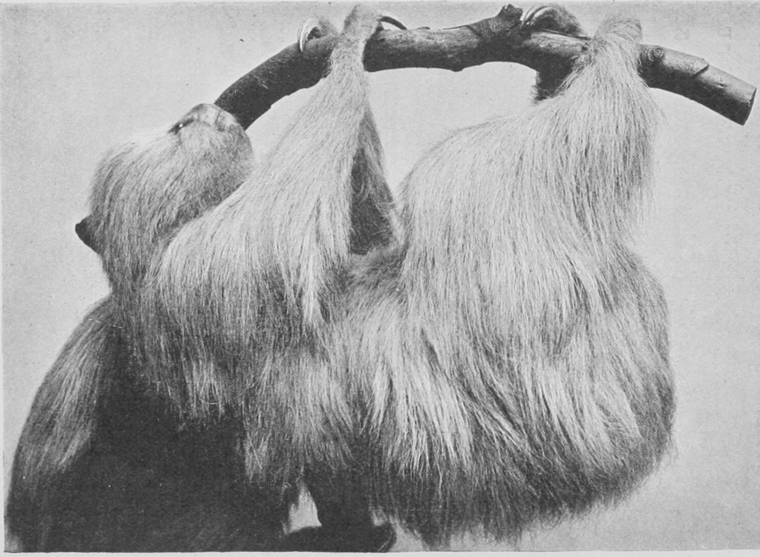
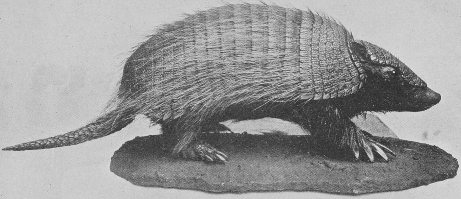
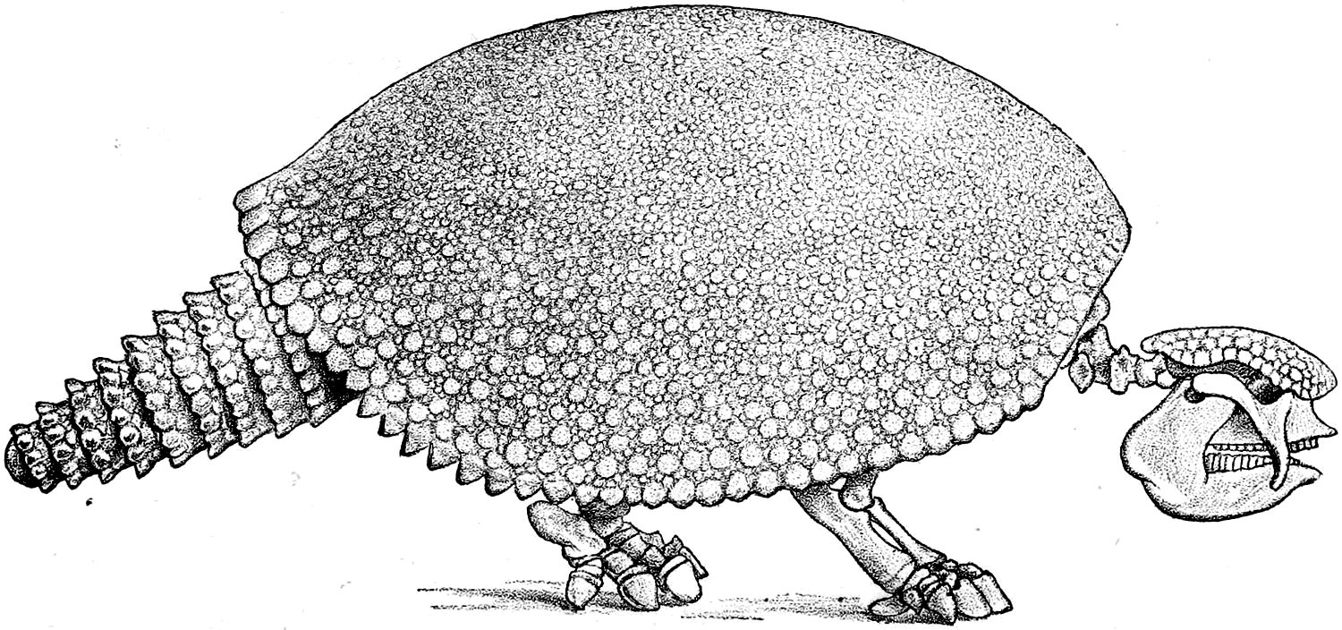
The Glyptodons (Fig. 120), of which there are several different kinds, were enormous armadilloes, as big as an ox. Like the recent little armadilloes they carried a hard case formed by bones in the skin, but this was not jointed so that they could roll up into a ball, as can the living armadilloes.
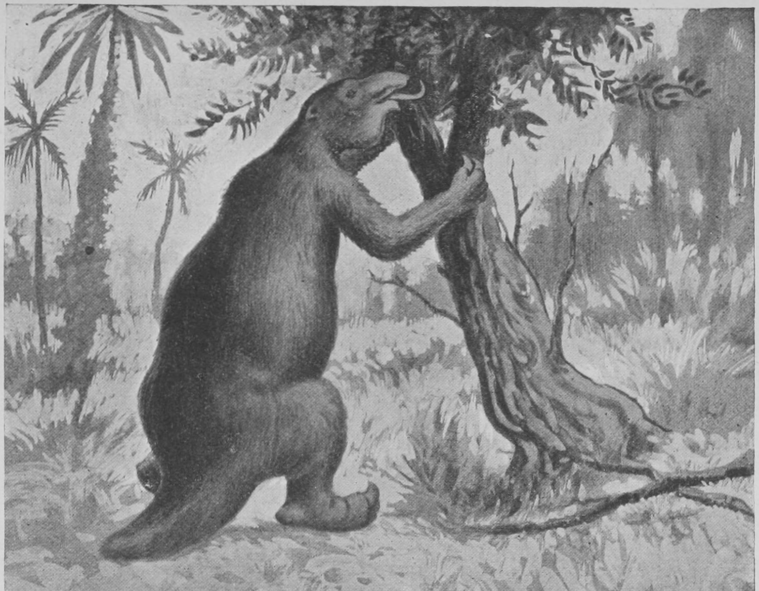
The Megatherium (Fig. 121) was nearly as big as an elephant, and was very closely similar in its skeleton and teeth to the little living sloths of to-day. But it stood on the ground and pulled the trees down in order to eat the tender young branches instead of climbing up into the trees and living there as the present sloths do.
Not quite so big as the Megatherium was the Mylodon, which lived at the same time. The remains of both are found in the comparatively recent (Pleistocene) gravels of the Argentine Republic. The skeletons of these animals may be seen side by side in the Natural History Museum.
In Fig. 122 is represented the skeleton of the Mylodon, and just above it, for comparison, is placed the photograph of the skeleton of the two-toed sloth. The relative sizes of the two are shown and the sloth's skeleton is placed in the same position as that of the extinct Mylodon, although in life it is always hanging from the branches of trees and never goes on all fours on the ground.
The Mylodon had, we know, a number of little bony pieces scattered in its skin in the region of the back, like the pieces forming the bony case of the armadilloes and Glyptodons, but not fitted closely together. It was supposed that the Mylodon, like all the gigantic Edentata of South America, had long ceased to exist and was extinct as long ago as the mammoth and the woolly rhinoceros of our own country. But about seven years ago a traveller (Dr. Nordenskjöld) found in Patagonia, at the end of a fiord near the Chilian coast, a vast cavern (Fig. 123), and from this cavern the white settlers living in a farm close by had removed an enormous piece of skin (Fig. 124) covered with greenish-brown hair and studded on its inner side with little knobs of bone (Fig. 125)! The skin was dry, but undecomposed, and when soaked in water gave out the smell of decomposing animal matter. It was evidently a piece of the skin of a Mylodon which had survived in this region until modern times! Further explorations were made in the cavern by Dr. Moreno, of the Museum of La Plata, and by other persons, and as a result an immense quantity of bones were obtained and more portions of the skin of Mylodon with the hair on. The cavern had been inhabited probably several centuries ago by Indians, and human bones as well as "forks" made out of dogs' bones (Fig. 126) were obtained. The remains of as many as twenty Mylodons have been obtained from the cavern, and many of the bones have been cut or broken by human agency, the inhabitants of the cave having fed upon the Mylodons and split the bones to obtain the marrow! Some of the Mylodon bones, skulls, jaw-bones, leg-bones, etc., are smeared with blood and the soft tendon and membrane are still attached. The cartilage at the end of the long bones is still in place, dried and cracked in the drying. Not only that, but great balls of dung were found made up of the remains of masticated grass, indicating that the Mylodons lived in the cave. Moreover, a very large quantity of cut grass was found in the cave, and it has been surmised that the Indians kept the Mylodons alive in the cavern and fed them with hay brought in from the outside. Specimens of these objects and of others to be mentioned below are now in the Natural History Museum, and some idea of their number and variety may be formed from the photographs reproduced in Figs. 126 to 131.
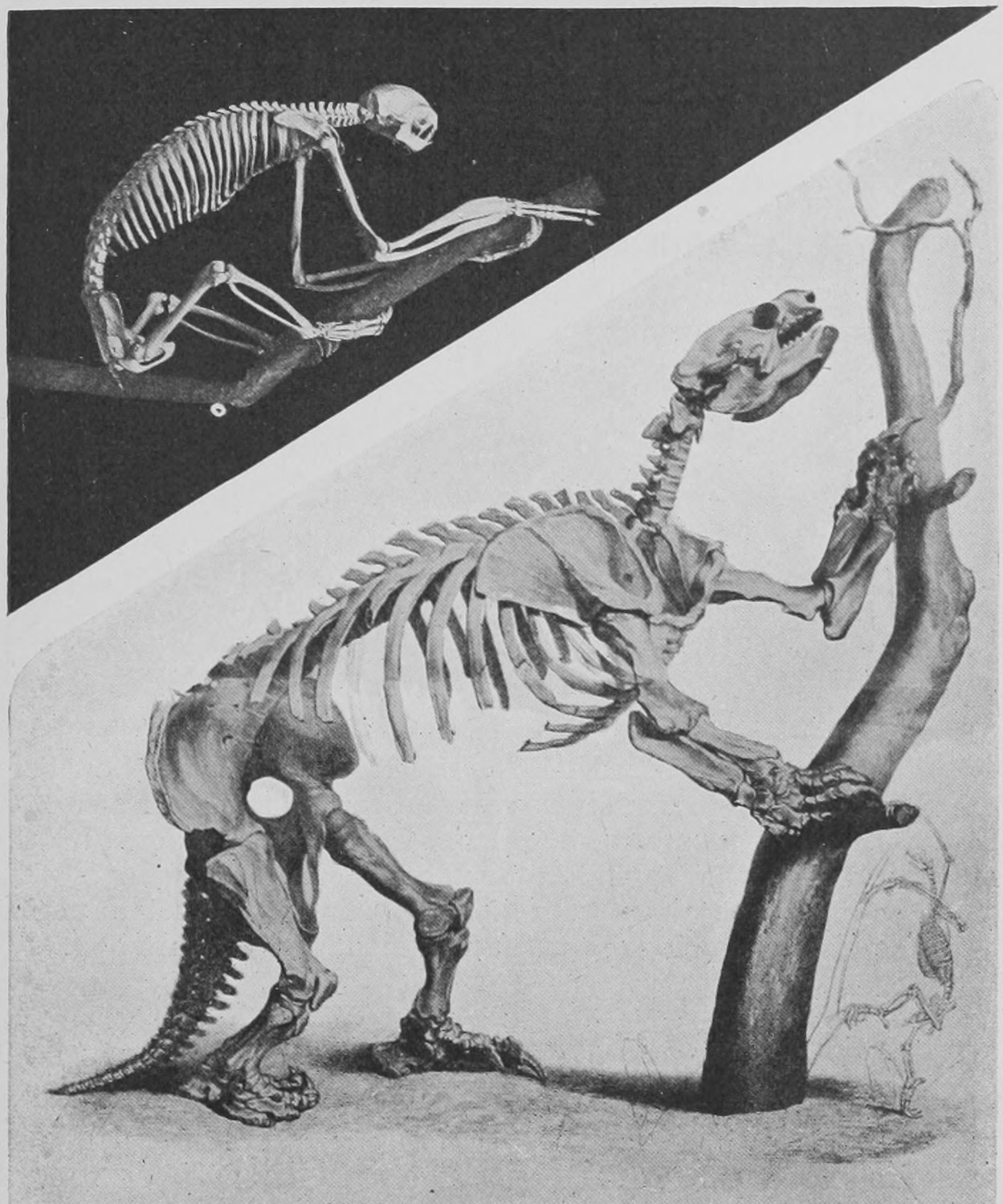
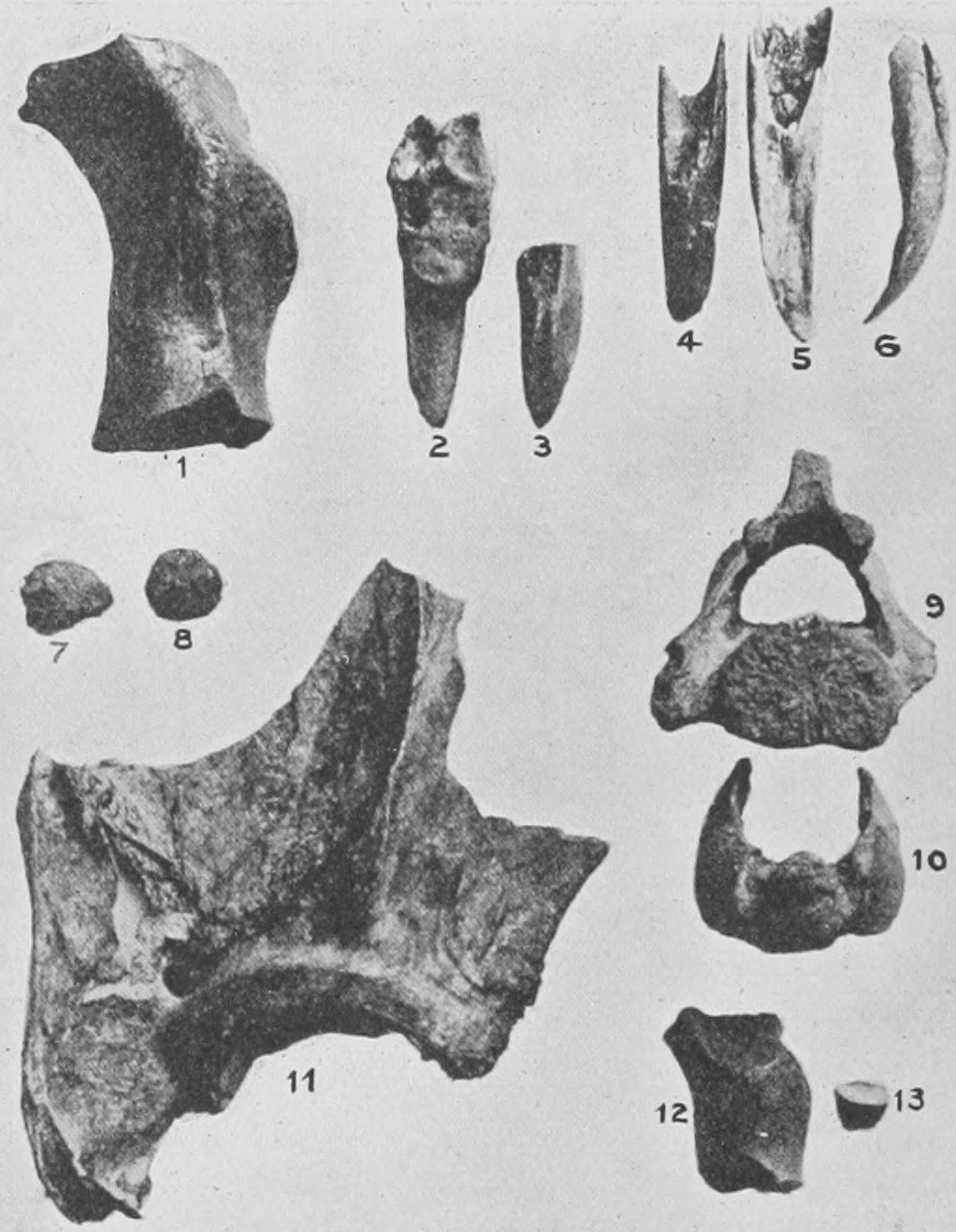
Besides the remains of the Mylodons and of man—all lying loosely covered by a greater or less depth of blown sand, and in some parts by chopped hay—the cavern has yielded bones and teeth and many horny hoofs of horses, apparently belonging to the extinct and very peculiar South American genus Onohippidium, the skull and bones of a very large kind of jaguar, the skull of a young lama, and bones of other kinds. We have not yet a full account of all that has been found in the cave, nor have the contents, unfortunately, been removed with sufficient care to enable us to say which were lying more deeply in the sand and which were at a higher level and therefore more recently living. The cavern is in a very remote spot and seems to offer some peculiar difficulties to explorers, for neither Sir Thomas Holditch nor Mr. Hesketh Pritchard, the latter of whom started for the purpose, succeeded in reaching it. It is stated that there are other caverns of a similar nature in the neighbourhood. A great peculiarity about the occurrence of the remains of animals in this cavern is due to the fact that it has a dry sandy bottom. The bones are not embedded in "stalagmite" as is the case in the bone-caves of England and France, and whilst they are quite unaltered and full of animal matter, the horny and tendinous parts of many of the animals, such as skin, hair, claws and hoofs, and the soft dung of the Mylodon, are preserved unchanged. It is quite certain that in any known cavern in Europe such remains would be destroyed in the course of fifty years by putrefactive bacteria, and were the conditions too dry for that process to continue, the remains would have been consumed by scavenger beetles and other insects within the like period. The climate of South Patagonia, where the cavern exists, is similar to that of Devonshire. It is a moist climate, although the cavern itself is not damp nor subject to inundation by streams. There is nothing in the sandy soil of a preservative nature, and it seems at first sight impossible to suppose that the soft dried remains, skin, claws, blood, etc., can be more than fifty years old. Yet the horses' hoofs and bones seem to belong to the extinct Onohippidium, and there is no record or tradition among the present race of Indians (in spite of some statements to the contrary) of any huge beast corresponding to the Mylodon. Altogether the case is a very puzzling one, and excites a very eager desire for further exploration. A noticeable fact bearing on the matter is that the whole of the southern part of South America has been submerged rapidly and has rapidly risen again and is still rising at the rate of two feet a year in some parts, within the late Pleistocene period. Possibly the rocks and high lands where the Mylodon cavern occurs formed an island during the submergence where a number of individuals of the earlier fauna took refuge and survived until the re-elevation of the land, and so lived on in the present condition of the land surface until fifty or a hundred years ago. Possibly, though by no means probably, the Mylodon is still living in similar caverns in this region, as yet unvisited by man.
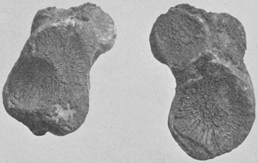
In Australia, the land of the marsupials or pouched mammals, the bones of gigantic creatures have been found belonging to that peculiar tribe. Giant kangaroos, twice as tall as any living kangaroos, are thus known. But there are also remains of some extraordinary animals, like wombats and koalas, only as big as the largest rhinoceros or a small elephant. One of these is the Diprotodon of Owen, known to him by its skull and the rest of the skeleton, excepting the feet. The skull is drawn in Fig. 132 with a human skull beside it to give a scale. In Fig. 133 is given Owen's restoration of the complete skeleton with the exception of the feet. These have now been found by Dr. Stirling, of South Australia. A number of complete skeletons of this huge beast were found embedded in the mud of a great lake or morass. In the photograph (Fig. 134) the lake is shown, and one of the great skeletons is seen in the foreground. The bones were in a very friable state, but Dr. Stirling has succeeded in preserving them and has secured the complete feet. In Fig. 135 the right hind-foot is shown. It is expected that the complete skeleton will be put together and exhibited in the Natural History Museum before very long.
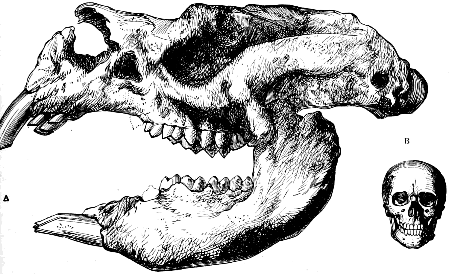
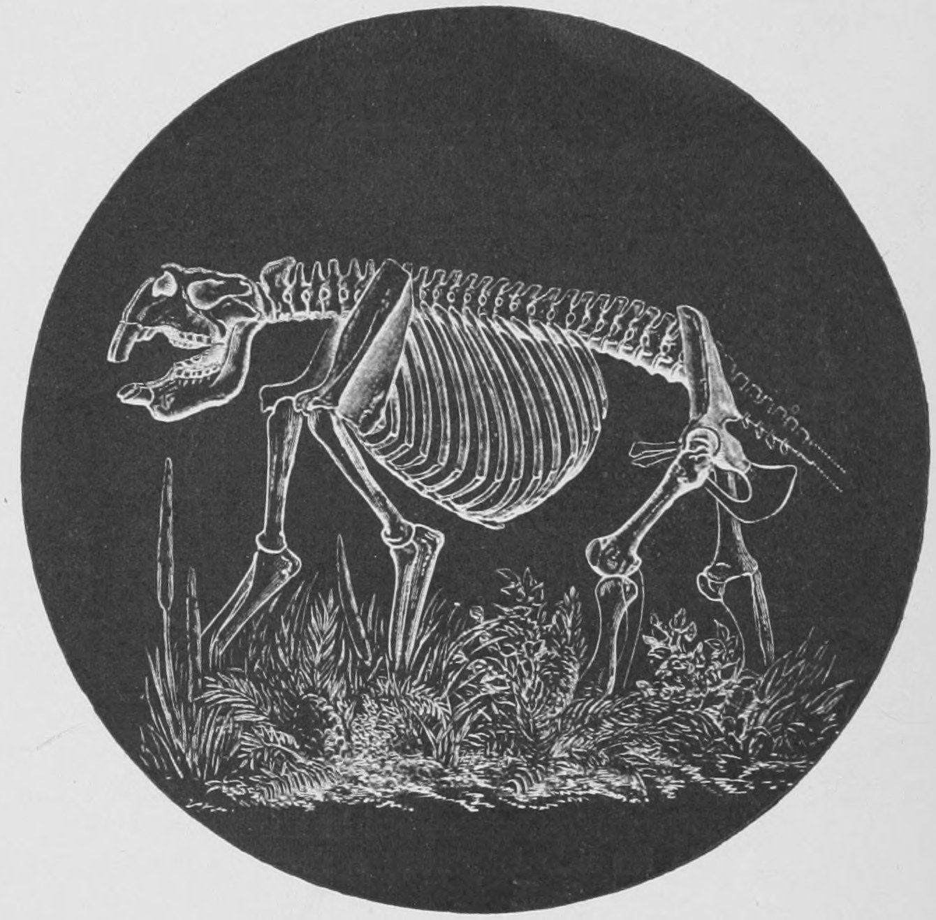
The oldest remains of mammals, which we know of, are found in the Oolitic and Triassic strata and consist of very small lower jawbones with their teeth, embedded in very fine-textured rock. It is usually held, on account of the form of the angle of the jawbones, that they belonged to small marsupial mammals. They are very small, few of them as much as an inch in length, and one of them we have already seen in Fig. 57 enlarged to ten times its natural length. It is probably due to their density and hardness that the little jaw-bones have been embedded and preserved in these ancient rocks, whilst the rest of the skeleton is lost to us. The first specimens of jaw-bones of this age were obtained seventy years ago in the Stonesfield Slate near Oxford by two undergraduates of the University, and it was at first supposed, on account of their occurring in such ancient rock as the Oolite (see list of strata on page 60) that they must be jawbones of lizards. Soon, however, the fact was noticed that the teeth had double fangs, and it became clear from this, as well as the shape of the jaws and teeth, that they had belonged to small mammals. In Fig. 136 two of these very ancient mammalian jaws are figured.
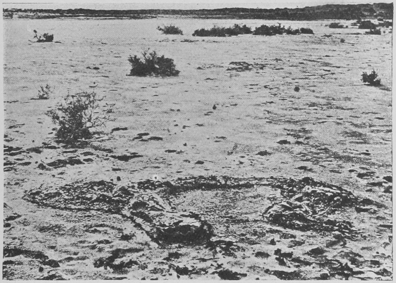
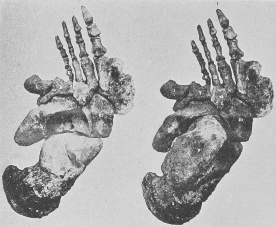
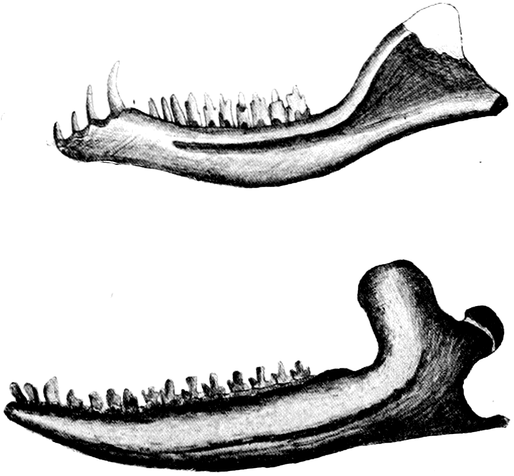
Chapter Five: The Great Extinct Reptiles—Dinosaurs from the Oolites—The Pariasaurus and Inostransevia from the Trias of North Russia and South Africa—Marine Reptiles
In the next two chapters I propose briefly to bring before you a few examples of extinct reptiles, birds and fishes, and to take the very shortest glance at the host of invertebrate shell-fish, insects, star-fishes and such like extinct animals whose name is legion.
We will proceed at once to the reptiles. You will see from the list of groups of reptiles which I gave to you in a former chapter (p. 58) that there are four big orders or groups of living reptiles: (1) the Crocodiles; (2) the Tortoises (Chelonians); (3) the Lizards; and (4) the Snakes. The lizards and snakes are in their real structure so much alike that they are considered as one double order. Extinct representatives of all these orders are found right away down through the Mesozoic strata to the Trias (see table of strata, p. 60). But there is nothing very astonishing about them excepting the large size of some of the extinct tortoises and snakes, and the fact that the older extinct crocodiles had the opening of the nose-passages into the mouth-openings, which we and all air-breathing vertebrates also possess, placed far forward as they are in the more primitive air-breathers, whereas living crocodiles have them pushed ever so far back to the very furthest recess of the long ferocious mouth, from which arrangement it results that the modern crocodile can have its mouth full holding the body of a victim under water whilst the air passes from the tip of its nose through the long nasal passage to the very back of its mouth and so to its lungs. This convenience was not enjoyed by primitive crocodiles.
The great interest in regard to extinct reptiles centres in those which were so entirely different from the reptiles of to-day that naturalists have to make separate orders for them. Many of them were of huge size. They flourished in the Mesozoic period and abruptly died out; at any rate their remains disappear from the rocks at the close of the Chalk or Cretaceous period (see the table of strata, p. 60). These extinct orders of reptiles are the Dinosaurs, the Theromorphs, the Ichthyosaurs, the Plesiosaurs and the Pterodactyles. They are a prominent example of that kind of extinct animal which is not the forefather, so to speak, of living animals, but of which the whole race, the whole order, has passed away, leaving no descendants either changed or unchanged.
To begin with the Dinosaurs. They are a very varied group and mostly were of great size. They seem to have occupied in many ways the same sort of place on the earth's surface which was filled at a later period by the great mammals, such as elephants, rhinoceroses, giraffes, giant kangaroos, etc. Preying on the vegetable-feeding kinds there were huge carnivorous dinosaurs, representing the lions and tigers of to-day. Yet the mammals I have mentioned are in no way descended from these great reptiles. They came from another stock, and only superseded them on the face of the earth by a slow process of development, in which the great reptiles disappeared and the great mammals gradually appeared and took their place.
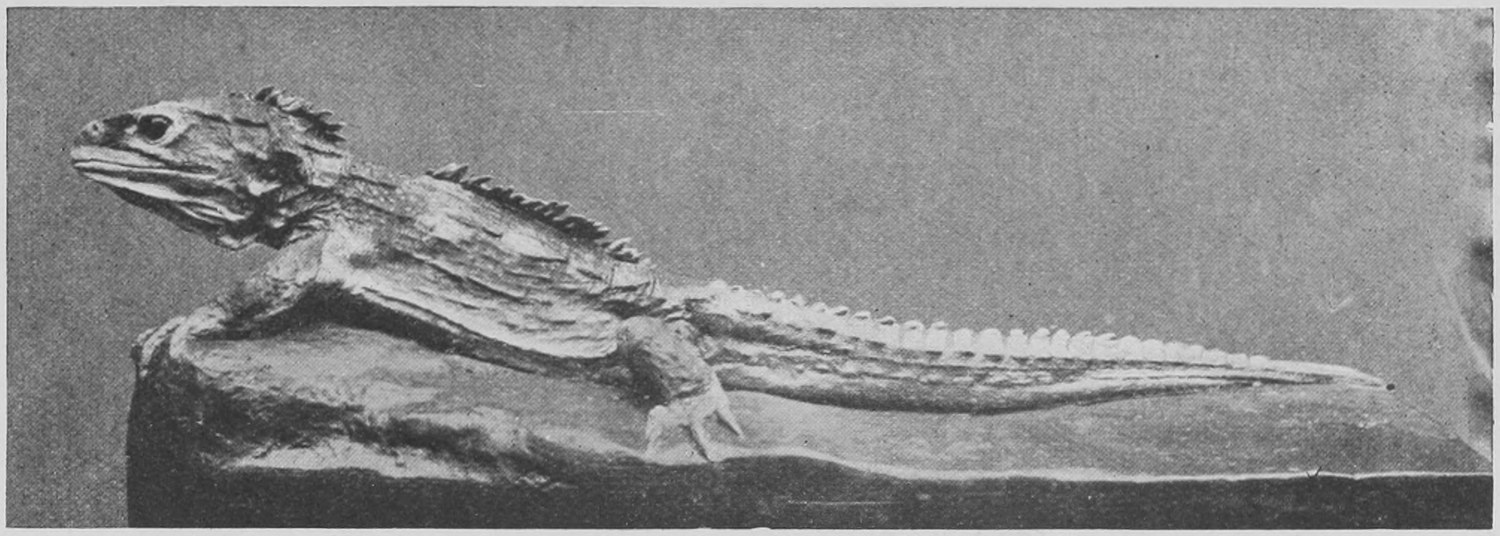
Some of the forms assumed by the great Dinosaurian reptiles are not unlike the forms of the small scaly lizards of to-day (see Figs. 137, 138, 139, 140); but on the whole the Dinosaurs were more like mammals in shape, standing well up on the legs. We do not know much about their skin; it was probably smooth and with only small horny scales on it, as in many living lizards, and often had great horns and crests growing out of it. But we know the complete skeletons put together from bones chiselled out of the hard rock in which they are found, and we know that in important matters of shape and build the skeleton was different from that of living reptiles. The great size to which some of the Dinosauria attained is shown by the thigh-bone of one found in the United States, and called Atlantosaurus—photographed in Fig. 6, p. 11. This thigh-bone is one third as long again as that of the biggest elephant known.
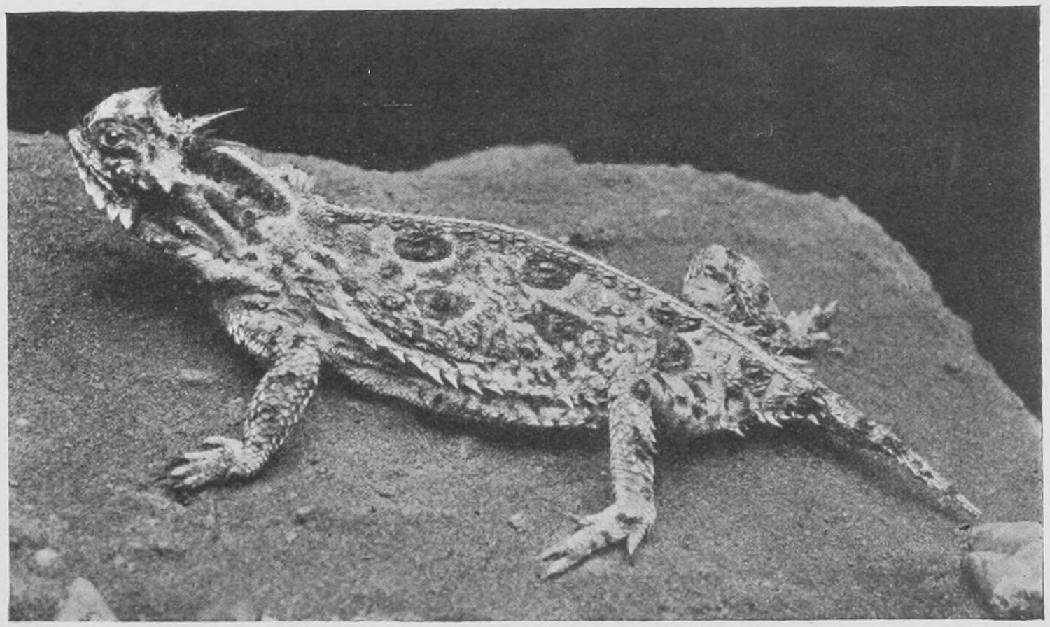
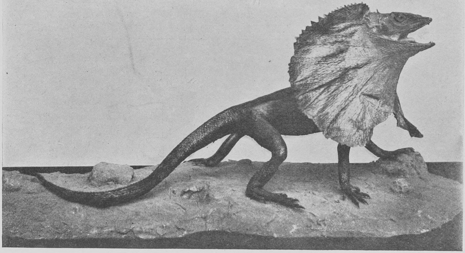
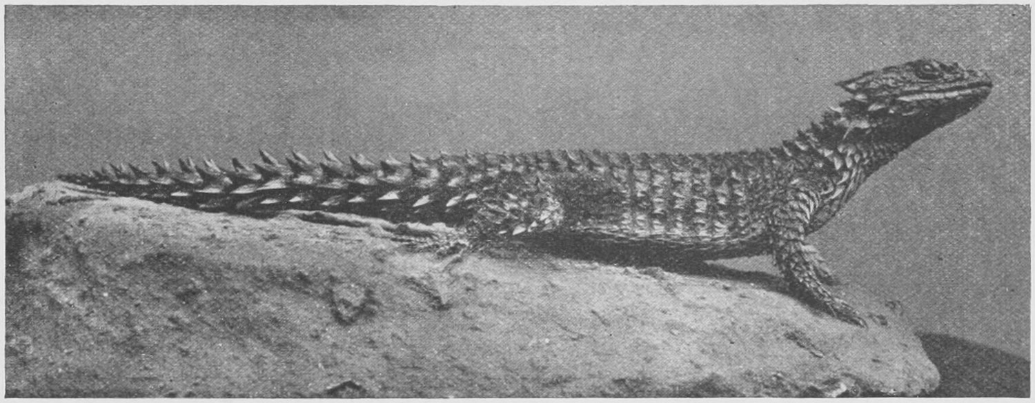
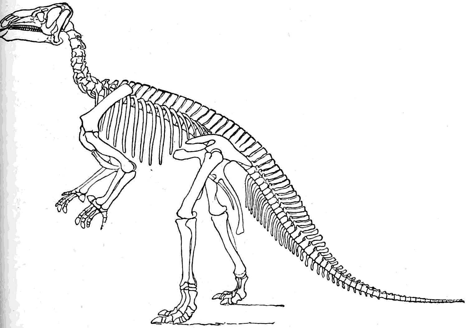
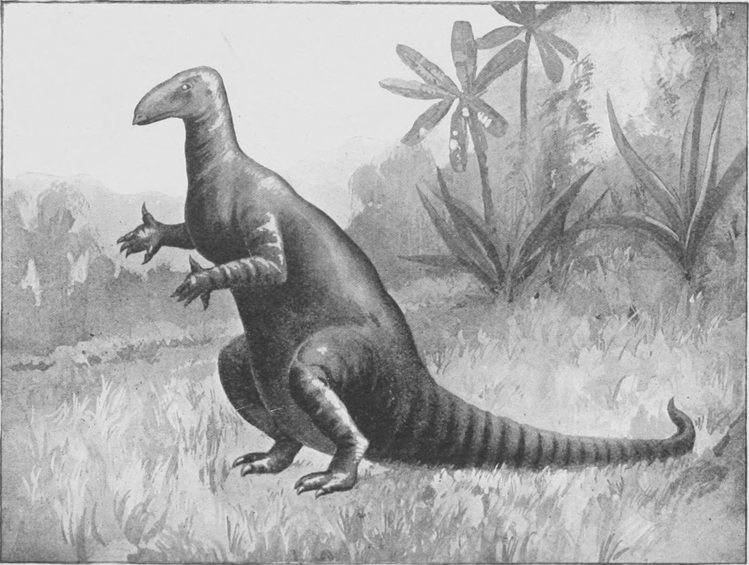
In Fig. 141 is shown the complete skeleton of the Iguanodon. This great Dinosaur was one of the first to be discovered. As you see, it stood on its hind legs like a kangaroo, and in running occasionally went on those feet only, touching the ground now and then with its front feet. Footprints in slabs of sandstone, once soft wet sand, are found showing this. The animal stood about fourteen feet from the head to the ground in the position shown in the figure. Its thigh bone was only three feet long and it was therefore only half the size, in linear measurement, of the Atlantosaurus.
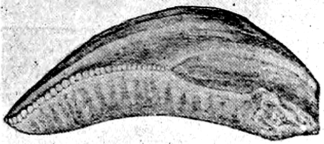
In Fig. 142 an attempt is made to show what the animal looked like when the skeleton was clothed with flesh and skin. The first bones and teeth of the Iguanodon were found seventy years ago by a celebrated and most delightful collector and explorer of the earth's crust, Dr. Gideon Mantell, in the strata known as the Wealden in Sussex, just below the Chalk and Greensand (see table of strata). Dr. Mantell found that the teeth, of which two are here represented of the natural size, were those of a herbivorous animal and like those of the little living lizard from South America, called the Iguana, in the fact that the broad chisel-like crown has a saw-like edge (Fig. 144). From this fact the name Iguanodon (Iguana-toothed) was given to the new fossil giant reptile. The bones found by Mantell and others were scattered and not in their natural position and the form of the creature had to be guessed at by fitting this and that together. But some twenty-five years ago a wonderful find was made near Brussels in a coal-mine at a village called Bernissart. The skeletons of no less than twenty-two huge Iguanodons were found complete, and embedded in a fairly soft clay-like rock! The authorities of the Government Museum took charge of the place and most carefully removed the rock containing the skeletons to the Museum workshops at Brussels, where the complete skeletons of seven were, with enormous difficulty and care, removed bit by bit from the rock and set up as entire skeletons in the Brussels Museum, where they may be seen. A cast of one of these seven is in our own Natural History Museum. The photograph of the skull of one of these specimens is given in Fig. 145. It shows not only the teeth in position, but in front the bony supports of a great horny beak, like that of a turtle. As you may see in the drawing of the skeleton (Fig. 142), the forefeet (or hands) were provided with five fingers, of which the thumb had a huge claw on it at least a foot long. The foot was very much like that of a bird and had only three toes, and the bones of the pelvis or hip-girdle are extraordinarily like those of a bird. In fact it is now certain that reptiles similar to the Iguanodon were the stock from which birds have been derived, the front limb having become probably first a swimming flipper or paddle, and then later an organ for beating the air and raising the creature out of the water for a brief flight. From such a beginning came the feather-bearing wing of modern birds.
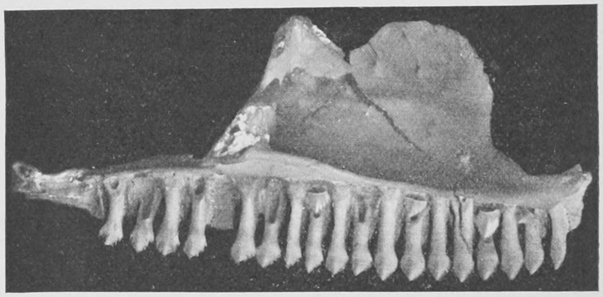
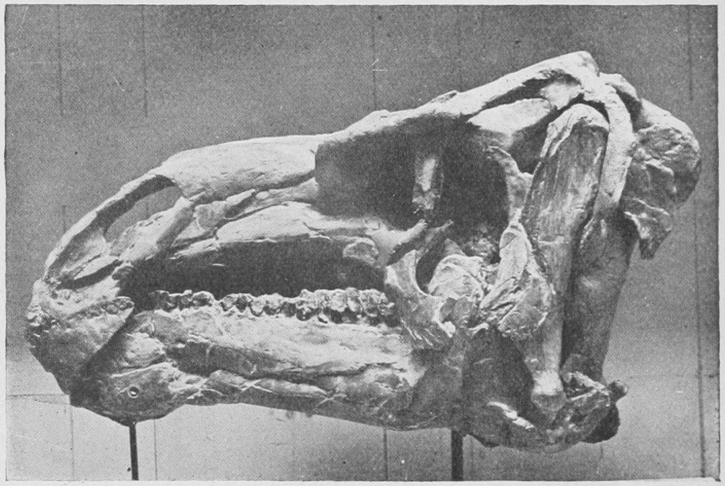
Fig. 146 shows the skeleton of a Dinosaur of somewhat less size but with the same kangaroo-like carriage, which was a beast of prey. It is the Megalosaurus, and had many tiger-like teeth in its jaws. It hunted down and fed upon the herbivorous Dinosaurs as lions and tigers hunt and eat antelopes and buffalo to-day. By no means all the Dinosaurs walked on their hind legs. There were enormous kinds which went on all fours. Here is the skeleton of the Brontosaurus (Fig. 147) and a sketch of its appearance in life (Fig. 148). The great Ceteosaurus, of which the limb bones and most of the skeleton were found near Oxford, is similar to this, and Mr. Andrew Carnegie has presented to the Natural History Museum a complete reconstruction of the skeleton of a closely allied Dinosaur—the Diplodocus—which was excavated in Wyoming and is now in the Carnegie Institute at Pittsburg. It is eighty feet long. Its head is very small, and a great part of the length is made up by the very long neck and the very long tail, but the body is bigger than that of the biggest elephant and the back was nearly fourteen feet from the ground.
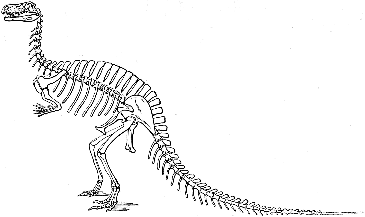
The immense profusion in which the bones of these huge creatures have been found in Mesozoic strata in the United States is astonishing; no less remarkable is the skill and success with which American naturalists—chief among whom have been Professor Marsh of Yale and Professor Cope of Philadelphia—have collected, fitted together and drawn every detail of more than thirty different kinds of these monsters. They have given such full evidence of the structure and build of the animals that we may with confidence accept the reconstructions of the appearance of the animals such as those shown in Figs. 149 and 150, where the rhinoceros-like Triceratops and the huge crested Stegosaurus are represented. Such crests and horns are bizarre and grotesque even when carried by little living lizards a few inches long, but it must be remembered that the Dinosaurs drawn in Figs. 149 and 150 were as big in the body as large elephants.
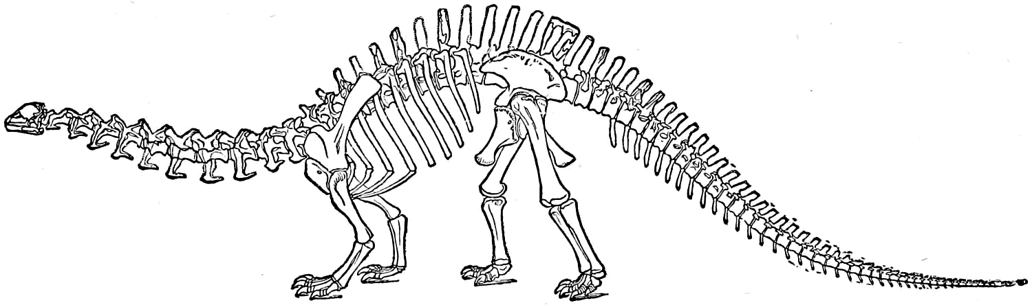
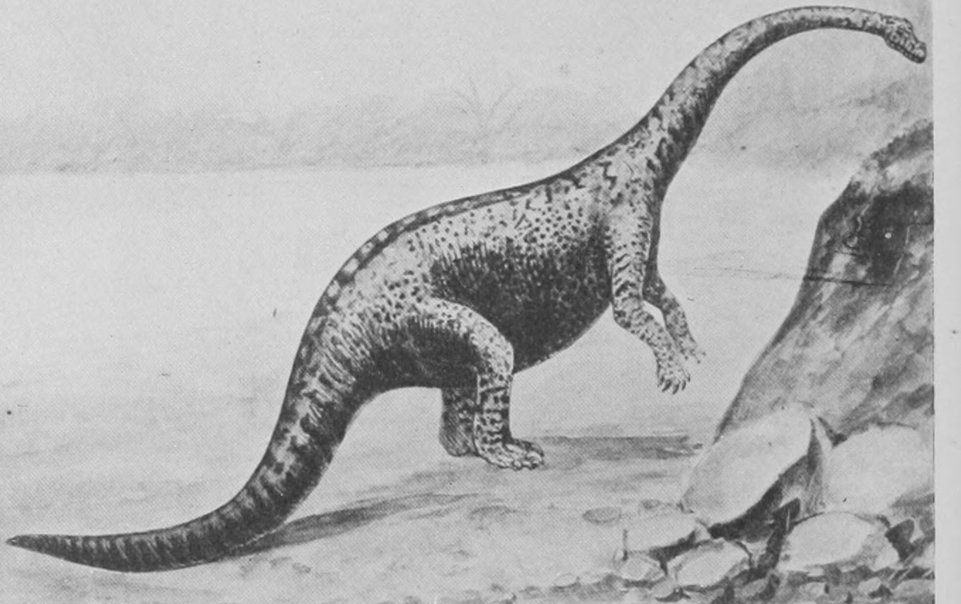
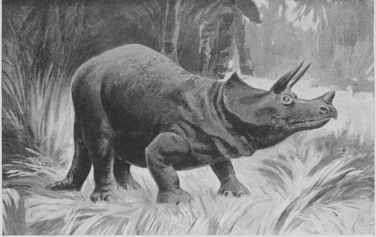
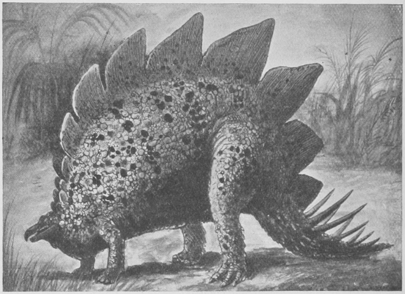
A curious fact about these great Dinosaurs is that they had, as compared with big living reptiles such as the crocodiles, very tiny brains. You will remember that the extinct mammals known as Titanotherium and Dinoceras have brains one-eighth the bulk of living mammals of the same size, such as rhinoceros and hippopotamus. So it was with the huge extinct reptiles. In some the head itself was ridiculously small according to our notions of customary proportion, and even in others, such as Triceratops, where the bony and muscular parts of the head were big, as in a rhinoceros, yet the brain was incredibly small. It could have been passed all along the spinal canal in which the spinal cord lies, and was in proportion to bulk of body a tenth the size of that of a crocodile. Very probably this small size of the brain of great extinct animals has to do with the fact of their ceasing to exist. Animals with bigger and ever increasing brains outdid them in the struggle for existence.
So much for the Dinosaurs, which might well occupy a complete course of lectures all to themselves. We will now turn to the Theromorphs, which are an older group even than the Dinosaurs and flourished in the Trias period (see table of strata, p. 60). The Theromorphs are so called because in some important parts of the structure of skull and jaw, and often also in the teeth, they resemble the mammals or Theria. They come near to a point in the history of terrestrial vertebrate beasts which is the common origin of Reptiles, Mammals and Batrachia or Amphibians (newts, salamanders and frogs).
Their remains have been found in the Triassic sandstones and limestones of South Africa, of Russia, of India and of Scotland and the centre of England. One of the most striking of these is represented by a completely reconstructed skeleton from Cape Colony in the Natural History Museum, photographed in Fig. 151. The skeleton is some eight feet long and looks like a gigantic pug-dog. This is the Pariasaurus, and is shown by its small teeth to have been herbivorous.
From the same locality we have the Dicynodon with two huge tusks, and the Cynognathus with a skull and set of teeth wonderfully recalling those of a bear at first sight.
Another strange crested form belonging here is the Dimetrodon from the Permian strata of Texas, U.S.A. (Fig. 152).
But I am now able to show you, through the kindness of Professor Amalitzky, of Warsaw, a set of photographs taken by him, showing the discovery and working out by him of a whole series of skeletons of these Theromorph reptiles, closely similar to those from the rocks of Cape Colony but belonging to a locality far removed from South Africa, namely, to the banks of the Northern Dwina near Archangel in North Russia. Professor Amalitzky has not yet finished his excavations nor published these photographs, and it is therefore a great kindness on his part to allow me to show them here in London.
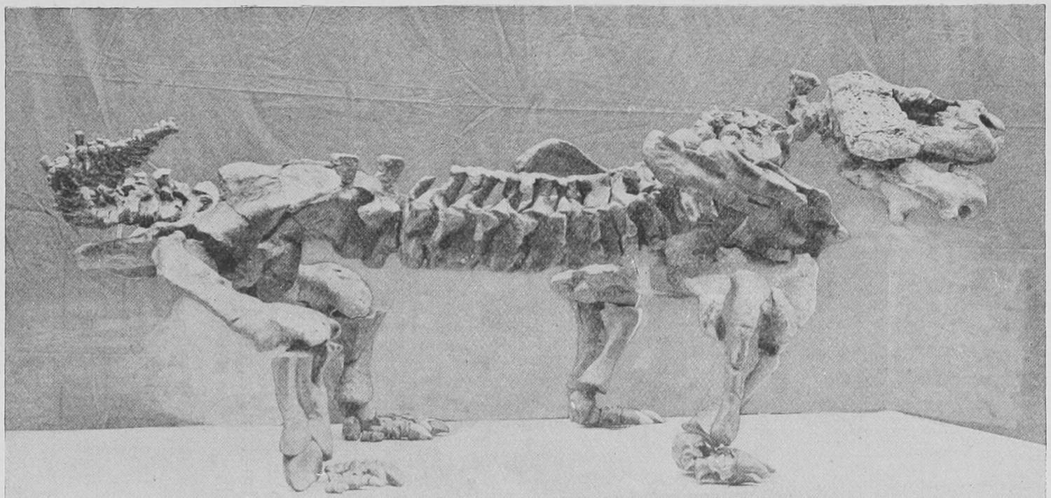
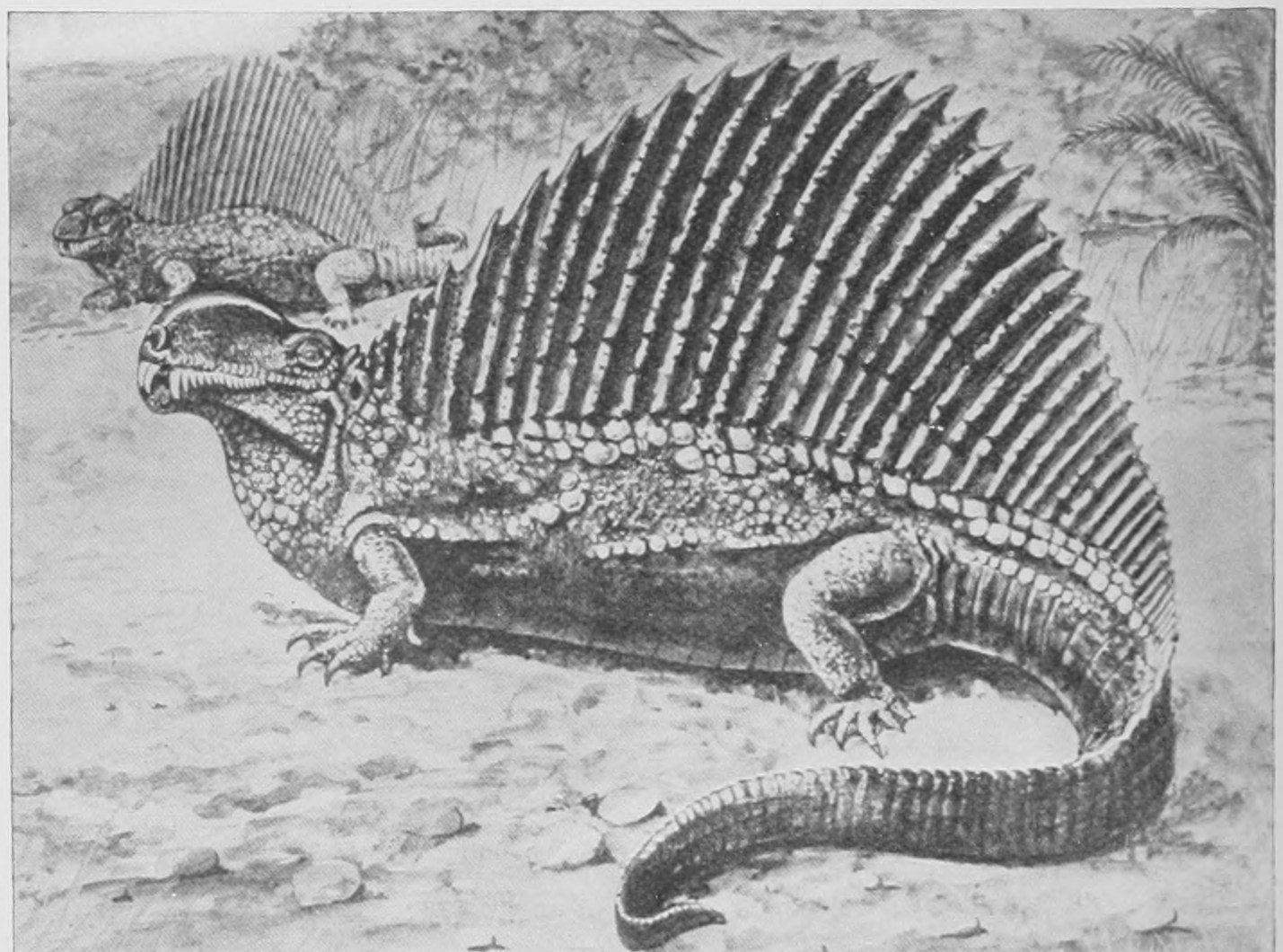
First of all, we have the cliff of Permian strata on the banks of the Dwina (Fig. 153), from which and from another similar spot the remains were extracted. At this point, where the colour is dark in the photograph, there is a peculiar "pocket" or accumulation of sandy matter with large hard nodules embedded in it. These nodules are removed and broken up for mending the roads. The pocket seems to be in a fissure and of Triassic age, later, that is to say, than the Permian rocks on each side of it. However that may be, the great nodules are removed from it for road mending, and four or five years ago Professor Amalitzky on visiting the spot was astounded and delighted to find that when broken each nodule was seen to contain the skeleton or skull of a great reptile. Fig. 154 shows such a nodule, some eight feet long, and in this specimen one can easily distinguish the skull, the four limbs and the backbone of a large animal. The Russian geologist determined to make a most thorough investigation of this wonderful deposit, and for some years now has spent a thousand pounds a year, obtained for the purpose through the Imperial Academy of St. Petersburg, in having the nodules dug out by the peasants after their farming work is over for the year, and in removing them to the University of Warsaw, where with the finest instruments and greatest care the nodules are opened and each bone removed in fragments is put together from its more or less broken parts, firmly cemented and set up in its natural position and relations as part of a complete skeleton.
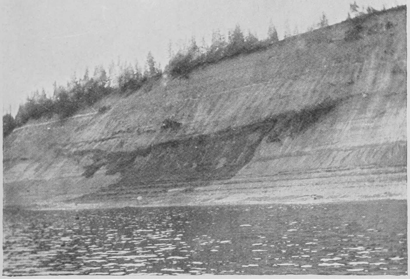
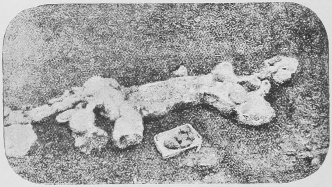
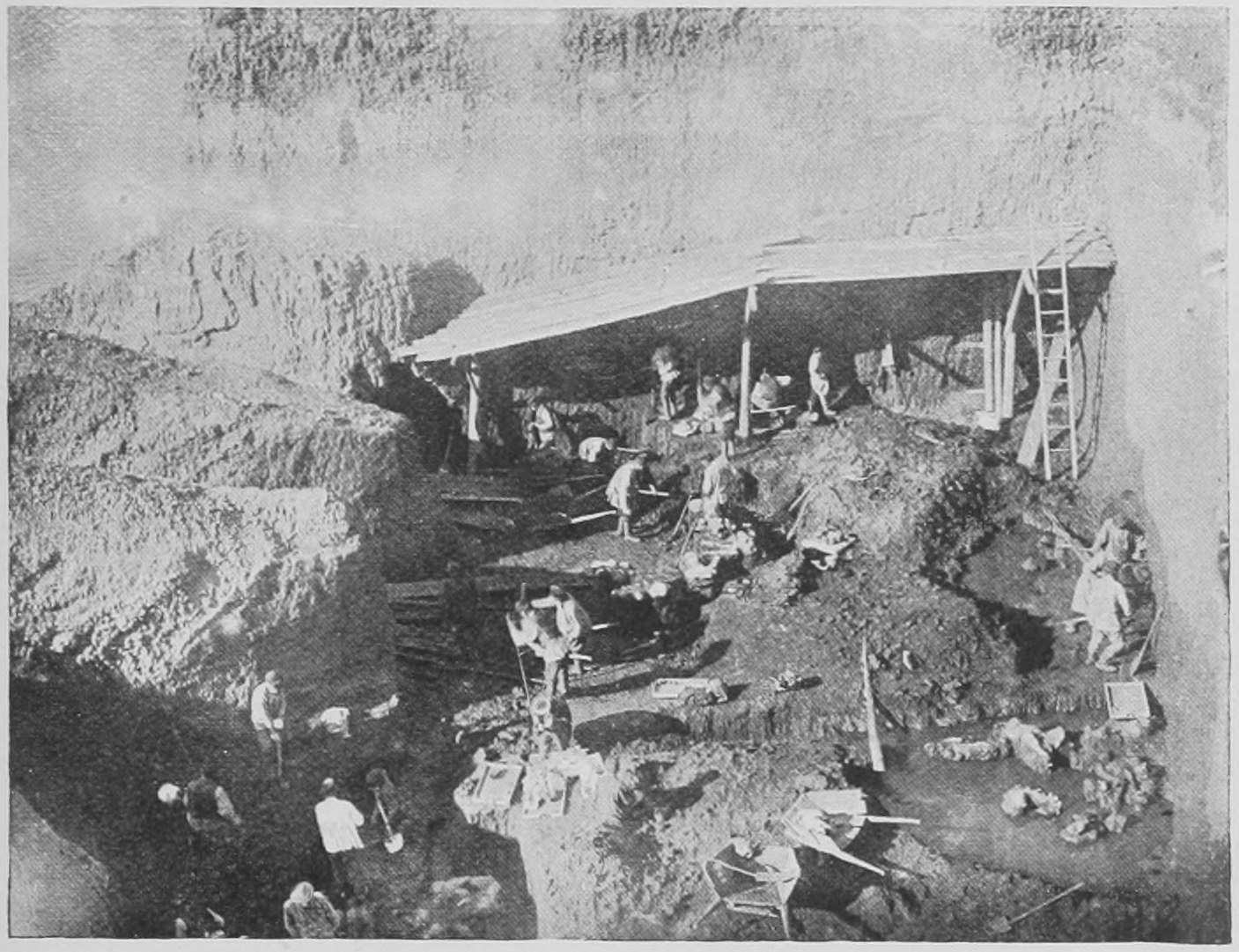
Fig. 155 shows the peasants at work, protected by a shed from the fall of stones from above. Fig. 156 shows some of the nodules as yet unopened lying in the laboratory of the geological professor at Warsaw. Fig. 157 shows a number of skeletons of the huge but harmless vegetarian Pariasaurus which have been cleared out of the nodules and set up on iron supports, as more or less complete specimens. Of course it is not possible in every individual to get out all the bones complete, especially those of the feet. Few of the individuals were complete even when originally embedded in the mud ages ago. When an animal's body is carried away by a river and floats in a decomposing state it tends to fall to pieces.
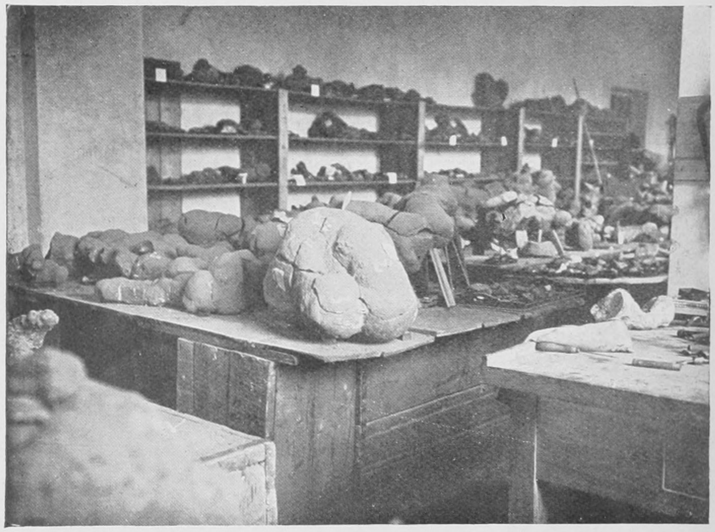
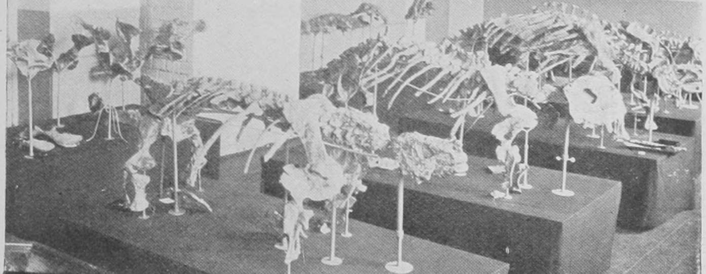
The cliff formed by the present river Dwina consists of rocks of immense, indeed of almost inconceivable, age, and existed as solid rock ages and ages before the surface of the earth had its present form. These deep-lying rocks have been brought near to the surface by bending of the strata (as shown in Fig. 36, p. 52), and the cutting or cliff made by the comparatively modern river exposes them to our view and to easy excavation. The nodules are relatively to the age of the river-valley or cutting (which is probably some 150,000 thousand years old), as much older than it is as are Roman coins older than the trench dug three hours ago which brings them to light. If you look at the position of the Trias and Permian in the table of strata you will get some idea of how immensely remote is the time when these great reptiles lived where now is Archangel, for whilst the thickness of a twentieth of an inch suffices to indicate the accumulations of strata since the mammoth lived in England, the Trias is a long way down the series, far below the Eocene, where the ancestral elephants of Egypt are found, far below the Chalk, and older than the long Jurassic series of rocks in which the remains of the great Dinosaurs we have recently looked at, occur.
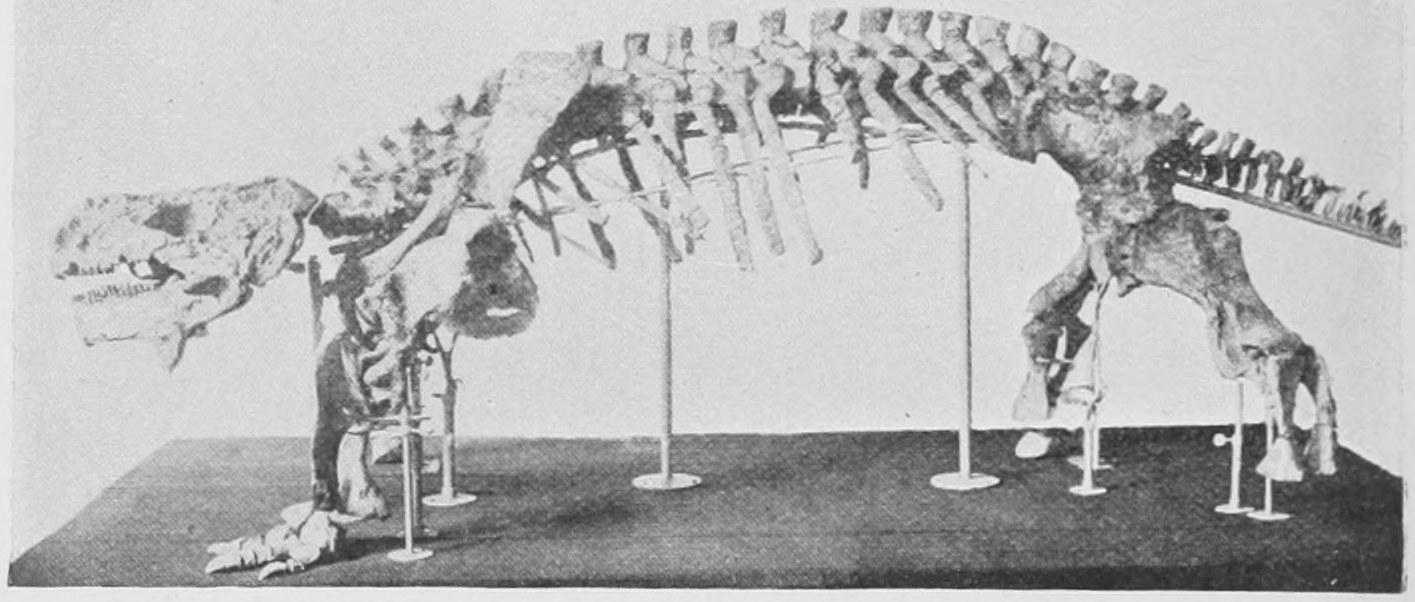
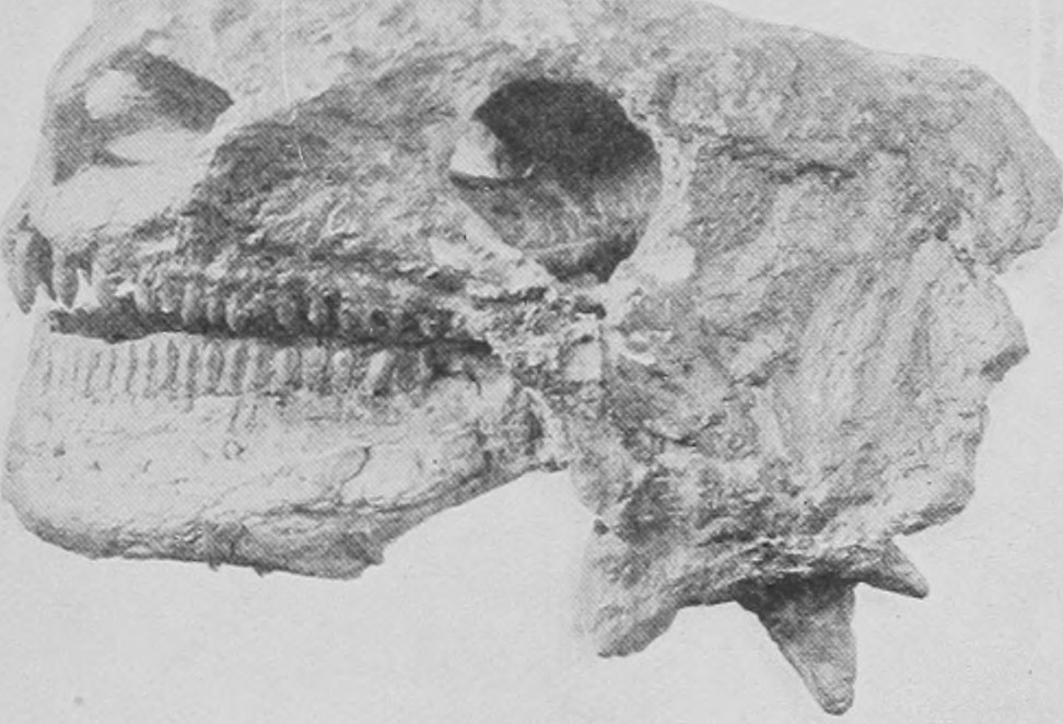
In Fig. 158 one of Professor Amalitzky's specimens of Pariasaurus is shown. There is no artificial completing of this skeleton: all that is seen is actual bone as cleaned out of a nodule. Only one foot is preserved, but that of course tells us as to its fellow of the opposite side. The skull of another specimen of Pariasaurus is shown in Fig. 159. It is very remarkable that this species seems to be so closely similar to the one discovered far away in South Africa in beds of the same age.
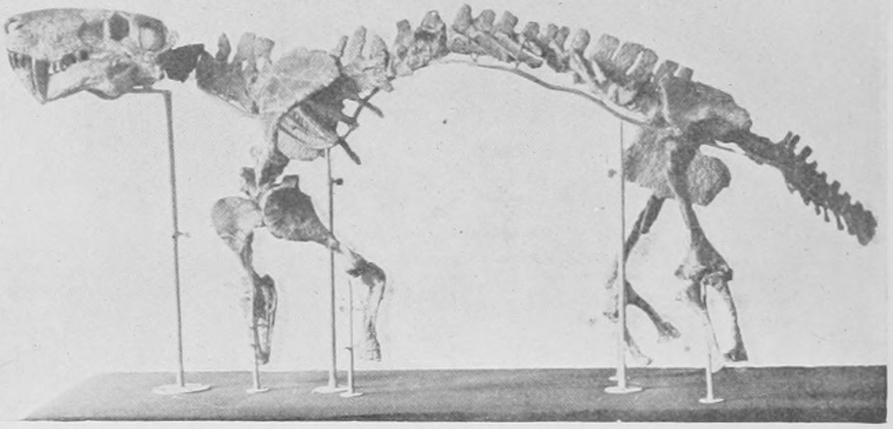
These Pariasaurs were about as big as well grown cattle, but not so high on the legs. In Fig. 160 we have the skeleton of another creature revealed by these nodules. It is an enormous and truly terrible carnivor, with a skull two feet long and enormous tiger-like teeth. This creature is named Inostransevia by Professor Amalitzky, and is larger than any of the carnivorous reptiles from South Africa. Specimens of its skull are shown in the Professor's photographs reproduced in Figs. 161 and 162.
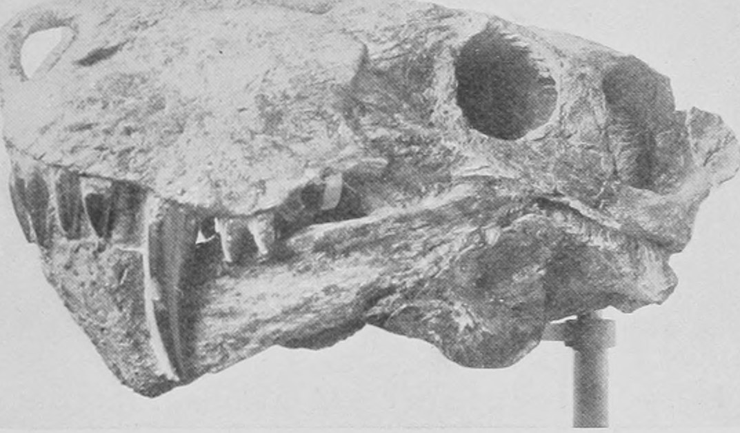
No doubt the vegetarian herds of Pariasaurus, whose small peg-like teeth indicate clearly enough their inoffensive habits, were preyed upon by the terrible Inostransevia, as were their brethren in South Africa devoured by the Cynognathus, the Lycosaurus, the Cynodraco and other carnivorous reptiles of that remote Triassic age. So we see the co-existence of blood-sucker and victim—of the destructive oppressor and the helpless oppressed—forced on our attention in these two localities, Russia and South Africa, when we study the immensely remote past of the Triassic age.
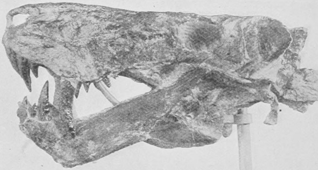
We leave now these great extinct land-dwelling reptiles and take a glance at representatives of two extinct orders of huge aquatic creatures which must also be classified as reptiles. These are the Plesiosauria and the Ichthyosauria. Though some of them must have measured thirty feet from snout to tail, they do not equal in size the great aquatic mammals of to-day, the whales.
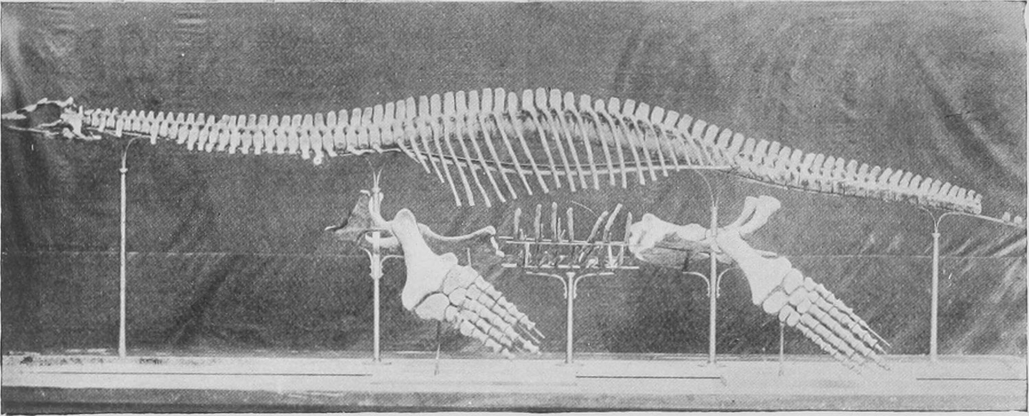
In Fig. 163 is shown the photograph of the skeleton of a large Plesiosaur, and in Fig. 164 is given a drawing showing how the creature appeared in life. It had a body like the hull of a submarine with four paddles attached, the fore- and the hind-legs. It had a long neck like that of a swan and an elongated head provided with powerful jaws armed with numerous pointed teeth. It probably could swim under water as well as on the surface, and when in the latter position could snap small lizards and birds from the land. The paddles have the definite structure of legs, with five toes, wrist or ankle and fore-arm or fore-leg and upper arm or thigh. A great number of kinds of these Plesiosaurs have been discovered, especially in the Lias rocks of the South of England, slabs containing whole skeletons being frequently obtained. They and the similarly embedded and flattened skeletons of different kinds of Ichthyosauria may be seen in quantity on the walls of the gallery of fossil reptiles in the Natural History Museum.
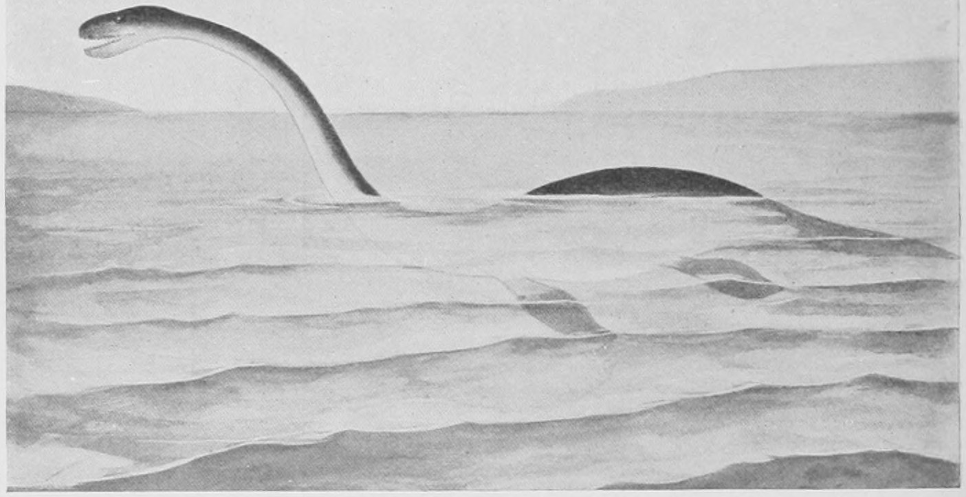
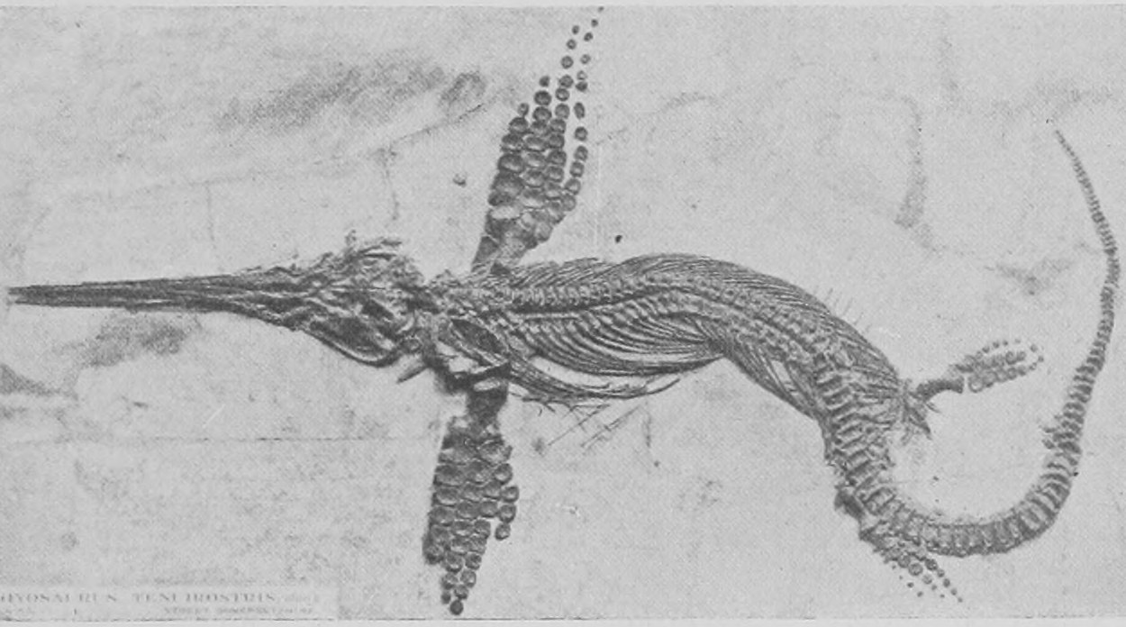
In Fig. 165 the flattened skeleton of an Ichthyosaurus is photographed. This particular species is remarkable for the great size of its fore-paddles.
In Fig. 166 a drawing of an Ichthyosaurus, as it must have appeared in life, is given. The Ichthyosaurs are much more fish-like or rather whale-like in form than the Plesiosaurs. They were indeed singularly like the porpoises and grampuses among living whales and stand in the same relation to land-living reptiles that the porpoises do to land-living mammals. Their fish-like appearance and fins are not primitive characters and do not indicate any closer blood-relationship to fishes than that possessed by other reptiles. They are the offspring of four-legged terrestrial reptiles which have become specially modified and adapted to submarine life. Like many whales they had a median fin on the back devoid of bony support. The bones of their legs have become greatly changed, much more so than those of the Plesiosaurs and form often more than five rows of nearly circular or polygonal plates fitted together as a flexible paddle. The tail is fish-like, but has the lower lobe bigger than the upper and the vertebral column bends down into the lower lobe instead of turning up into the upper lobe as it does in fish. The details as to the fins are known from some wonderfully preserved specimens found in the fine hardened mud known as the lithographic slate of Solenhofen, where the soft bodies of jelly-fish, cuttle-fishes and the wings of flying reptiles also are preserved.
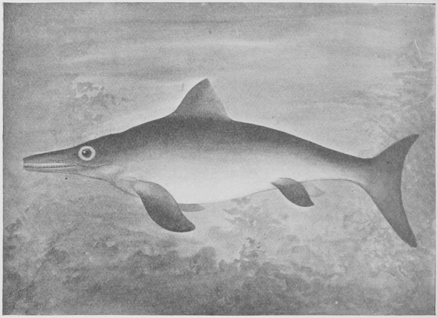
As mentioned in the first chapter, the Ichthyosauria (see Fig. 2) had a ring of bony plates supporting the eye-ball (as birds also have), and these are often preserved in the fossil specimens. In Fig. 168 a view of the top of the skull of an Ichthyosaurus is given in order to show the round hole in the middle line of the brain-case (on a level with the letter P). This is called the "parietal foramen," and is a fair-sized hole in which was lodged an eye, a third eye called the pineal eye. This eye is found in some other reptiles also, and especially in some of the living lizards where its structure has been studied with the microscope. There is no doubt that the body filling this hole in living lizards is an eye, although it seems to have lost the power of sight in these recent forms. A third eye, placed on the top of the head strikes one as a very strange arrangement and contrary to all our common experience of vertebrate animals.
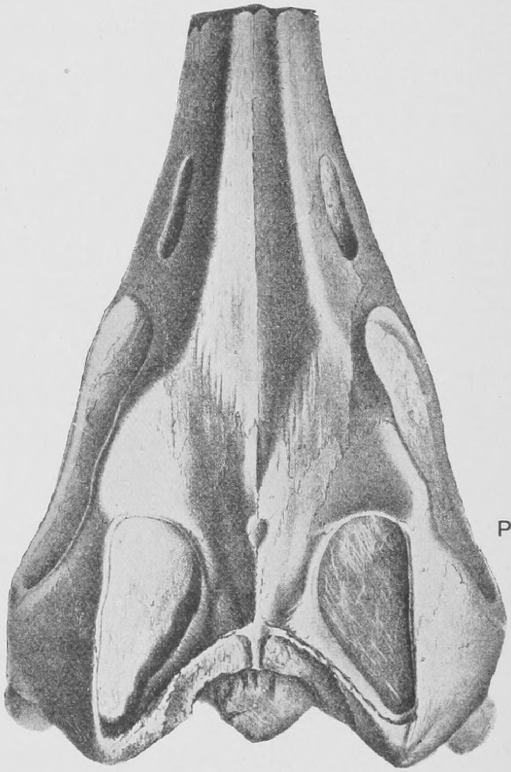

In Fig. 168 we have a drawing of the side view of the skeleton of an Ichthyosaurus and below it a fossilized lump of its excrement. These are called coprolites and consist of scales and bones of fishes digested by the Ichthyosaurus. They show a corkscrew-like moulding of the surface, proving that the intestine of the Ichthyosaurus had a spiral fold like a spiral staircase on the walls of the intestine, as have the sharks. We also find within well preserved specimens of the skeletons of Ichthyosaurus the skeletons of unborn young individuals, showing that the Ichthyosaurus brought forth its young alive.
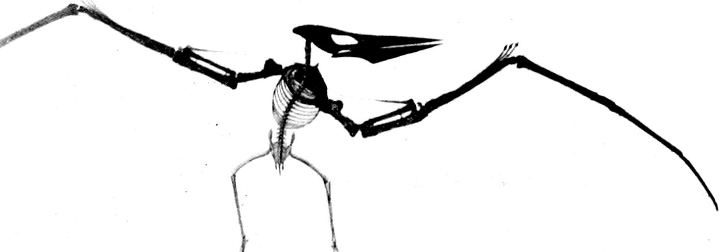
We pass on now to even more astonishing reptiles—the extinct order of the pterodactyles or flying reptiles of the Mesozoic period. These creatures were as truly aërial as the birds and bats of to-day. They were of many kinds, from the size of a crow to so huge a form as that drawn in Fig. 169, which measured eighteen feet from the tip of one wing to the tip of the other. Their wings have been found well preserved in the Lithographic slates (see Fig. 31, p. 47), and each consisted of a membrane spread from one enormously big elongated finger to the side of the body and little hind legs.
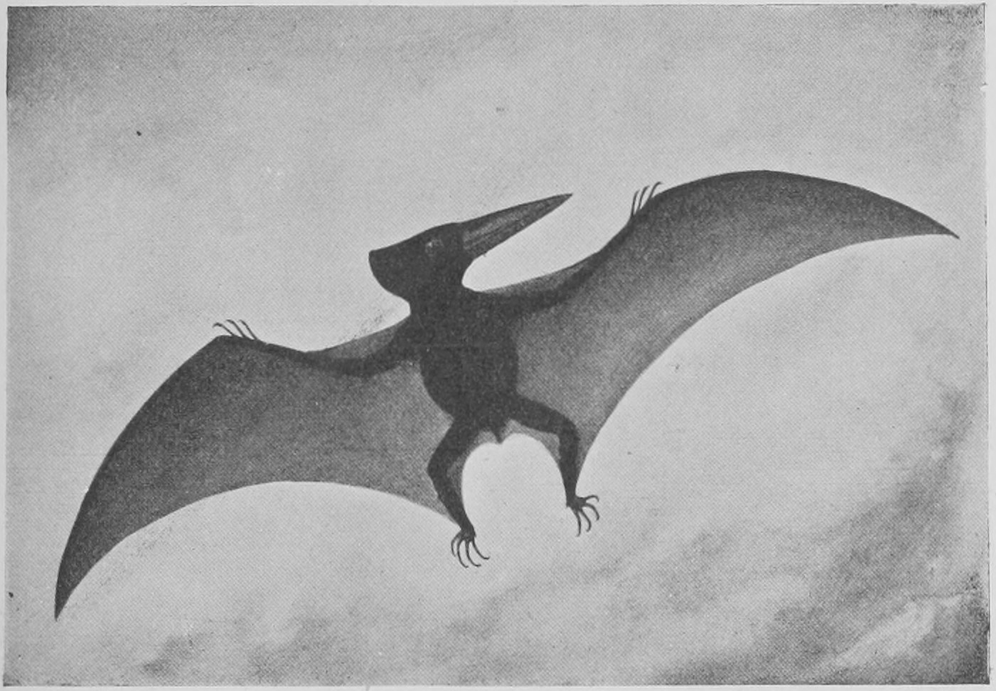
Fig. 170 gives some idea of the form and appearance of the wings when expanded. Such a wing is more like that of a bat than that of a bird, since it is a membranous skin and not a series of feathers. The bat's wing is a membrane supported by three of the fingers as well as the side of the body and hind leg.
In Fig. 171 the fossil wing of a Pterodactyle, that of a recent bird with the bones and the great quill-feathers only in place (the smaller feathers having been plucked off), and the wing of a bat are photographed and placed together for comparison. There are two other kinds of flying animals, namely, the flying fishes (which do not fly far), and the six-legged insects or flies, bees and beetles. They have all independently acquired the habit of flying and have had certain parts of their bodies changed into wings. The process of change must have been gradual and have taken an enormous lapse of time to bring it about in each kind. There are some animals, such as the flying squirrels and flying lizards (Draco volans) of to-day, which do not really fly, since they have no wings to beat the air with, but can spread out a great flat surface on each side of the body which enables them to sail through the air for some distance without falling when they jump from the branch of a tree. This, however, is a long way from the point reached by animals which have wings and can strike the air as a fish strikes the water with its fins. Probably the wings of birds and of insects were both derived from fin-like organs which were used to swim with—before they were used in the air. But the origin of the wing of the Pterodactyles, and independently that of the wing of the bats, does not seem to have been of this nature, and is one of the many very puzzling matters which further discoveries may one day enable us to understand.
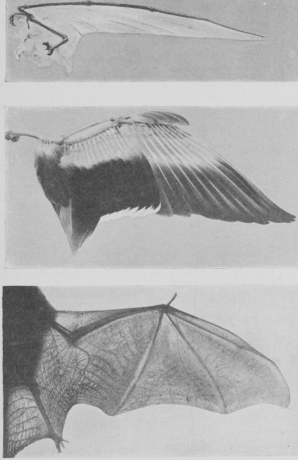
In Fig. 172 two other kinds of Pterodactyle are shown. Some Pterodactyles had no teeth, but long beak-like jaws (Fig. 169). Others had numerous sharp-pointed teeth and were beasts of prey.
It seems natural to pass from the winged reptiles to birds. But as a matter of fact the birds are not very closely related to Pterodactyles. Birds are, it seems, derived from reptiles, and are very specialized, warm-blooded descendants of certain reptiles. They are so peculiar that they are considered as a distinct "class," and the reptiles which come nearest to them in structure are the Dinosaurs, especially those Dinosaurs (like Iguanodon) which walked on their hind-legs and had only three toes to the foot. Fossil remains of birds are not abundant—but a few very interesting birds have been found in the Lower Eocene and in the Cretaceous rocks (see list of strata, p. 60), and one more remarkable than any other in the Lithographic slates of Jurassic age. Modern birds have all got feathers and beaks, and, with one or two rare exceptions, the quill feathers are set on the fore-arm and hand so as to form the wing. No living bird has teeth, but fossil birds are known with well developed teeth like those of reptiles. In Fig. 173 is shown the drawing of the skeleton of an extinct bird, which had a full set of teeth. The most remarkable extinct bird as yet discovered is that shown in Fig. 174. Two specimens of it have been obtained from the Lithographic slates of Solenhofen in Bavaria. The first one found is preserved in the Natural History Museum; the second and more perfect is in Berlin. This bird—called Archæopteryx—was of the size of a large pigeon, had a short head apparently without a beak, and its jaws were armed with teeth. Whereas living birds have the fingers of the hand aborted and tied together, this bird had three distinct fingers, each armed with a claw. Its legs were like those of living birds, and it had four toes. Its tail was unlike that of any living bird, and like that of a lizard. Whereas the bony part of the tail of living birds is very short and bears the tail feathers set across it fan-wise, the Archæopteryx had a long bony tail made up of many vertebræ, and the feathers were set in a series one behind the other on each side of it, so that the tail resembled the leaf of a date palm in shape. Strange as this little creature appears, it was a genuine bird, for it had true feathers well developed, which are clearly shown in the two fossil specimens. Besides the two rows of feathers on the long tail, there are the full set of feathers spreading from the fore-arms and hands to form the wings, and the thighs also were covered with feathers.
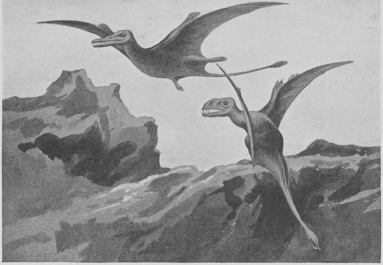
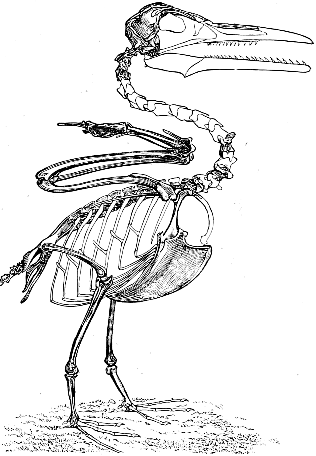
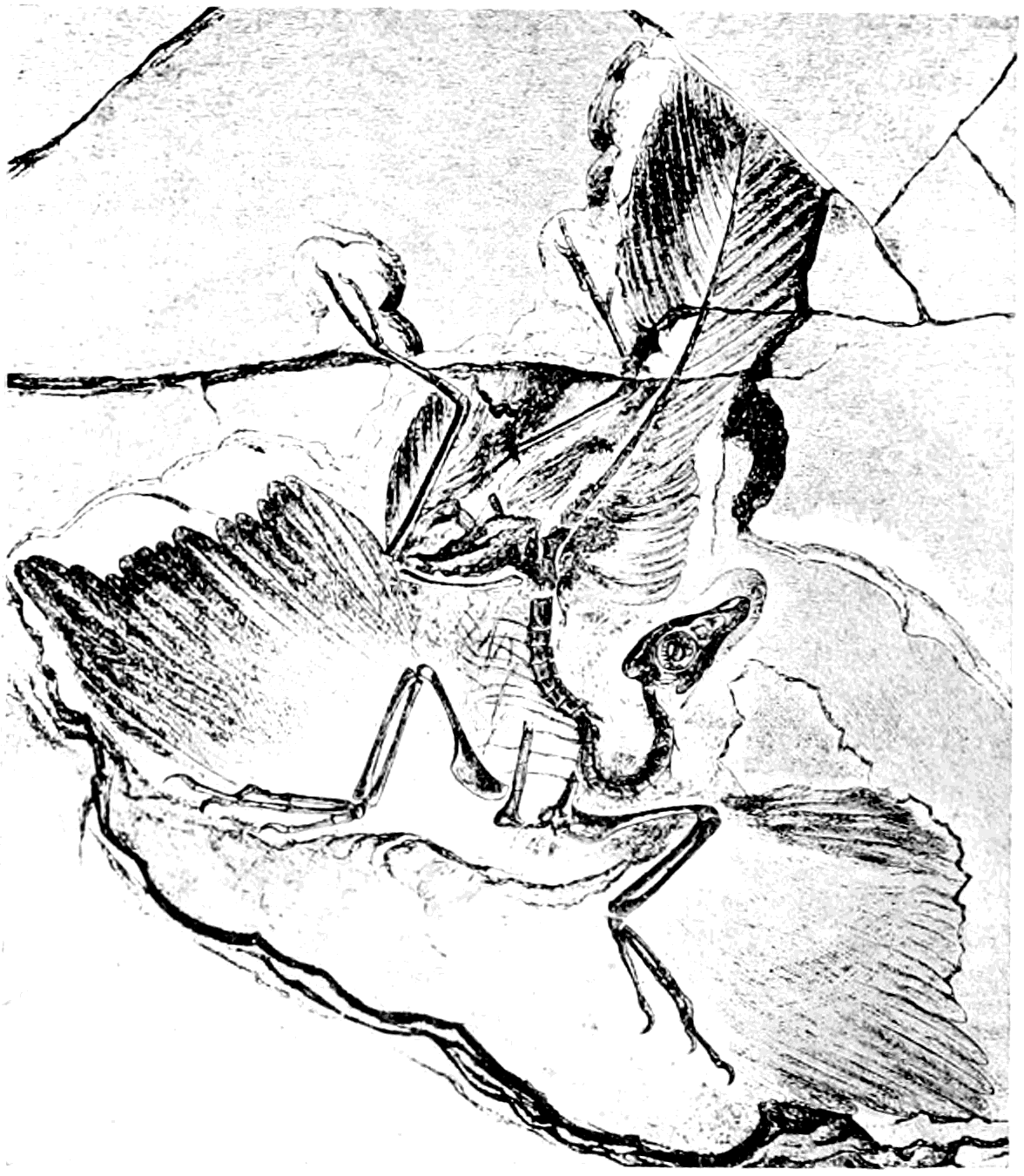
It cannot be said that this ancient extinct bird goes far towards connecting birds with reptiles: but in the possession of separate claw-bearing fingers, a long bony tail and teeth, in the apparent want of a beak, it does come nearer to lizard-like reptiles than does any other known bird.
In the Tertiary Strata remains of various birds have been found. One of great interest on account of its enormous size is the Phororachus of South America. We have in Fig. 175 a photograph of the skull of this bird placed beside the stuffed skin of a living South American bird, the Cariama or Screamer. If the extinct bird had the general proportions and habits of the Cariama, as seems probable, it must have been a terrible monster, standing some twelve feet high and far exceeding the most powerful eagles and vultures in strength and the size of its beak and claws. Great extinct wingless birds are found in quite recent "alluvial" deposits in New Zealand and in Madagascar. The discovery of the bones of the great Moa of New Zealand has already been mentioned in our second chapter (p. 69). Many species of Moa have been found in New Zealand. The Moa is allied to the ostriches of Africa, the emeus and cassowaries of Australia, and the rheas of South America.
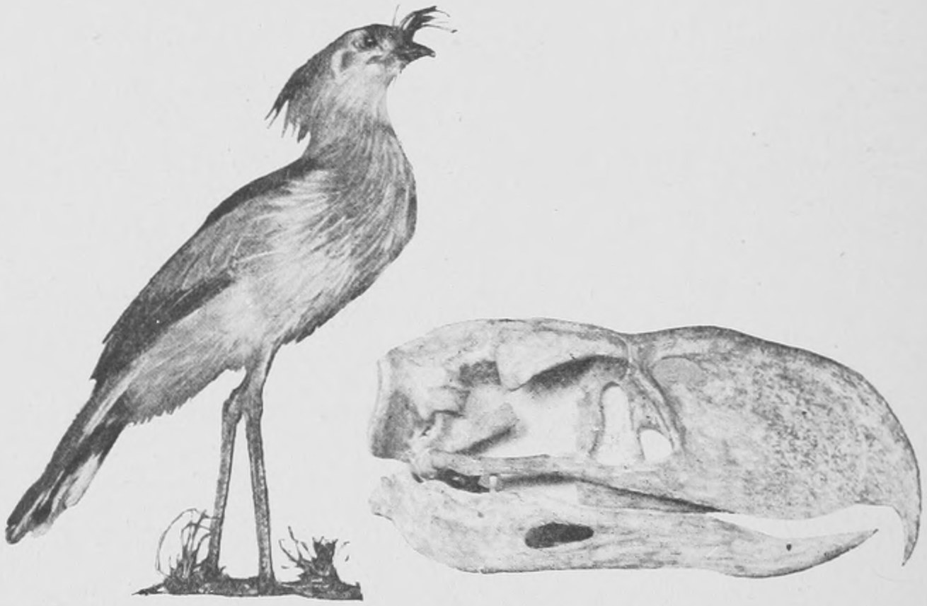
It appears that under certain conditions of life birds may gradually lose the use of their wings, which dwindle in size and finally may disappear altogether. Such wingless birds are not necessarily of one stock. The wingless condition, or the great reduction in the size of the wings, has occurred in various kinds of birds at various periods of the earth's history, and in the same way wingless insects of different orders have come into existence. In New Zealand, besides the Moas, which are all now extinct, a small kind of wingless bird is found which is still alive and is known as the Apteryx or Kiwi. In Fig. 176 we have placed one behind the other each with its egg in front of it: a Kiwi, the skeleton of a very fine Ostrich, and the skeleton of a giant Moa (Dinornis maximus). The Polynesian islanders who landed in New Zealand some five hundred years ago, found the Moas still living, and hunted them down and lived upon their flesh. Skin and feathers of these enormous birds have been found preserved in a dried condition as well as the skeletons, and there are traditions as to the hunting of the Moa still in existence. The Moa of Madagascar seems to have been a smaller bird, but laid a proportionately much larger egg. It will be seen in Fig. 176 that the eggs of the Ostrich and of the Dinornis are not nearly so big in proportion to the size of the bird as is that of the Apteryx, which lays a truly gigantic egg considering the size of its body. The Moa of Madagascar is known as the Æpyornis and laid the biggest egg known—much bigger than that of the biggest New Zealand Moa—resembling the Apteryx in the proportionate sizes of its egg and its body. It was this very large egg which inflamed the imagination of ancient navigators and led to the vast exaggeration, which thrills the reader with wonder and terror, in the accounts of the "roc" given by Sinbad the Sailor in the Arabian Nights.
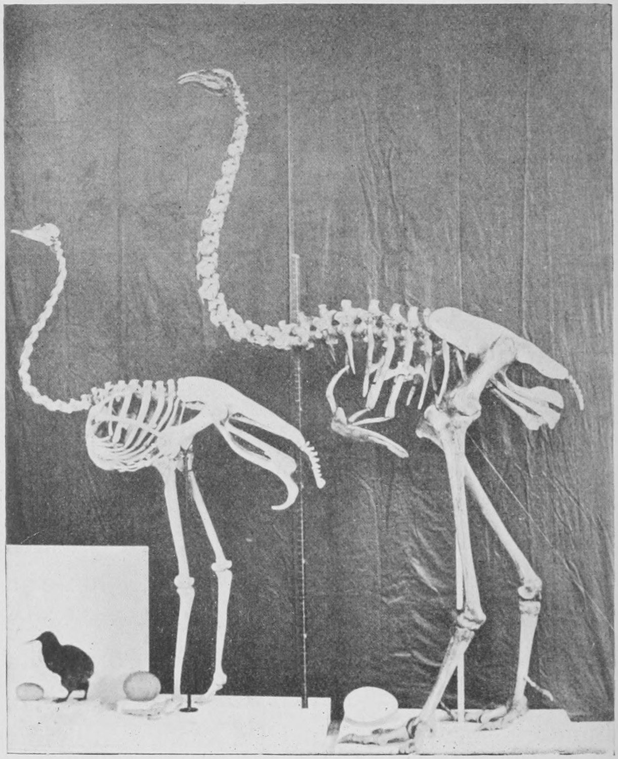
Flightless birds necessarily, unless they are, like the penguins, great swimmers, must get destroyed and become extinct when man arrives on the scene. The dodo, of which I spoke in my first lecture (p. 26), was a close ally of the pigeons, but had lost its power of flight owing to the fact that it had no dangerous enemies in the island of Mauritius. It had become a heavy, slow running, though powerful ground bird. As soon as man arrived, and with him the pig, the flightless dodo was doomed to extinction. An extinct water-bird, the Hesperornis, had no wings whatever, whilst the penguins use their wings as swimming organs and are unable to fly. This was also the case with the gare-fowl or great auk (Fig. 15, p. 23), which has recently become extinct.
Chapter Six: Extinct Fishes—Belemnites—Lingula—Trilobites—Scorpions and Stone Lilies
We might, if we had time, now look at the remains of the great bony Labyrinthodonts—creatures allied to the newts, salamanders and frogs of to-day, which form the class Amphibia. They stand lower than the Reptiles, Birds and Mammals; and though they have typically five toes and crawl or walk the earth, yet are essentially aquatic animals, inasmuch as their young are "tadpoles," fish-like in form and provided with gills. No reptile, bird or mammal has hitherto been found in what are called the Palæozoic strata, but in the Upper Palæozoic strata—those of the Carboniferous system, the period of the coal-bearing strata (see Table of Strata)—there was an immense variety of "Amphibia," some of very large size—as large as a well-grown crocodile.
It seems as though we might describe the Carboniferous as the period of the predominance of Amphibia, just as the Jurassic is that of the predominance of Reptiles and the Tertiary that of the predominance of Mammals.
The Labyrinthodonts, though of great interest to the trained anatomist, do not present many striking forms; the most noticeable were of the size and shape of large alligators. Accordingly, in the short space that remains to us, I propose to pass by the Labyrinthodonts and go on to the fishes, and bring to your notice some of the strange fishes, the remains of which are dug up in very ancient strata, as far back even as the Upper Silurian and the Devonian rocks. I shall then have space to mention a few of the more extraordinary extinct animals of the lower kinds, strange shell-fish, star-fishes and scorpions of the remote past.
The silver-scaled fish which are so abundant at the present day, with their symmetrical tails, such as herring, salmon, carp, roach, perch and other modern fishes more curious in form, such as eels, flat-fishes, sticklebacks, pipe-fishes and parrot-fish, are all of comparatively recent origin. They are not found in the rocks older than the Cretaceous system. On the other hand, the sharks and dog-fish of to-day are the most ancient kind of fish known to us, remains of shark-like fishes occurring in Silurian strata. But the sharks have soft cartilaginous skeletons, and have only, as a rule, left teeth and spines and the denticles of the skin (shagreen) in the rocks.
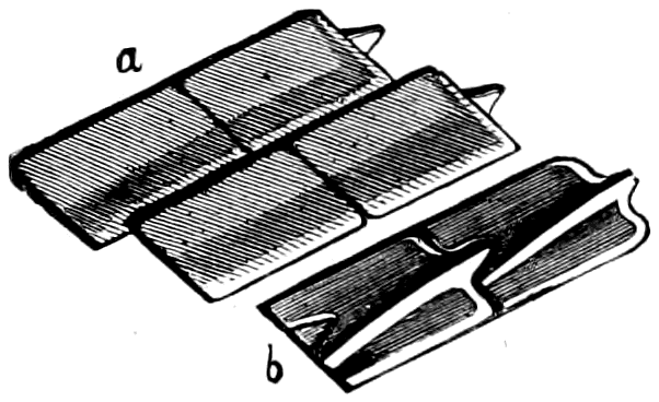
On the other hand there are certain fishes known which have hard bony scales and often great plates of bone on the head. They are often called "Ganoid fishes" on account of their hard smooth bony scales (see Fig. 177), which differ in substance and shape from the thin, flexible scales of common fish. They were very abundant in Mesozoic and Palæozoic times, and have left their hard scales in a very perfect state in the ancient rocks of those periods. They had often unequally divided triangular tail-fins, and in internal structure were like the sharks rather than the modern bony fishes. Very few of these Ganoid fish survive to the present day, but a fine one, the Polypterus (Fig. 178), still lives in the Nile and other African rivers, and another, the bony pike or Lepidosteus, in the North American lakes. The sturgeon also belongs to this set of fishes. In the Devonian is found, together with many others, a beautifully preserved fish, the Osteolepis (Fig. 180), which had lobed fins and hard bony scales like the Polypterus of the Nile. Allied to these Ganoid fishes, but differing in the fact that they possess lungs as well as gills and have very peculiar lobate fins, are the so-called mudfish of Africa (Protopterus) and of South America (Lepidosiren). A third mudfish is found in the rivers of Queensland, Australia, and is now living in the Zoological Gardens in London. It is called Ceratodus (Fig. 181), and is obviously related to some very ancient extinct fishes, of whose race it is a last survivor. One of these ancient forebears of the Ceratodus is found fossil in the Devonian or Old Red Sandstone of Scotland and of Russia. It is known as Dipterus, and is shown in Fig. 182. It differs from Ceratodus in having strong bony scales (whence its preservation as a fossil) and a triangular tail-fin. The true tail-fin has disappeared altogether in the living mud-fishes. Dipterus has peculiar teeth just like those of Ceratodus, and its fins are similar in character to those of the latter.
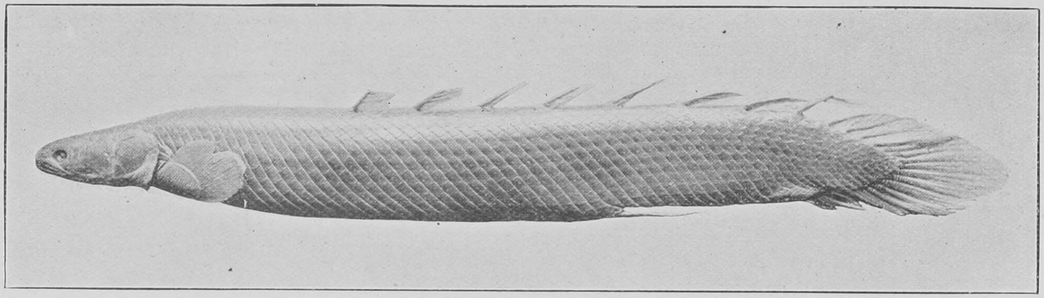
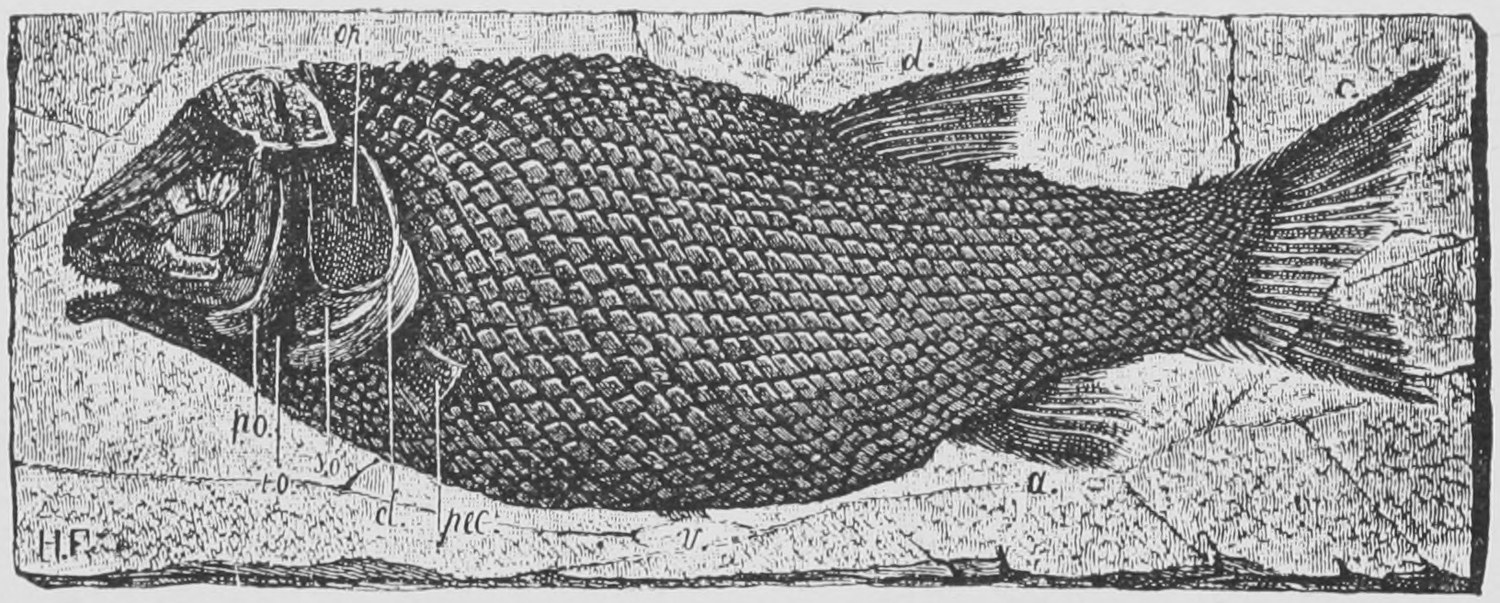
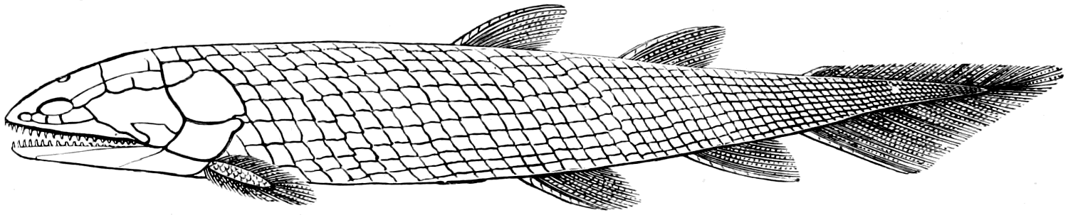
In the Devonian strata are found also the extraordinary fishes known as Pterichthys. They were compared by the wonderful Scotch quarryman, Hugh Miller—who seventy years ago discovered them and cleaned out many specimens from the rocks of his native hills at Cromarty in Scotland—to a tortoise's shield with a fish thrust into it. We have now gained from the examination of a great number of specimens from Canada as well as Scotland a very detailed knowledge of the curious bony plates which build up the case or "carapace" of the body of Pterichthys (Fig. 183), and also of its soft scaly tail, and the two extraordinary paddles or limbs which represent the anterior or breast fins of a common fish. Hugh Miller puzzled this out with great skill and constructed a cardboard model of the fish which we have still preserved in the Natural History Museum. It will, I think, be interesting to those who have read the writings of Hugh Miller (The Testimony of the Rocks, My Schools and Schoolmasters, and other books) to see a photograph of the model of Pterichthys which he made with his own hands (Fig. 184).
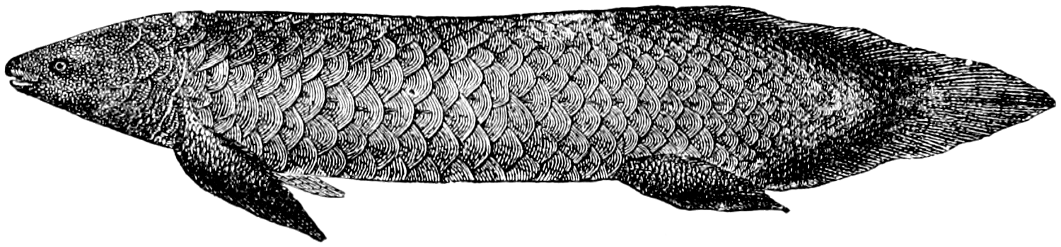
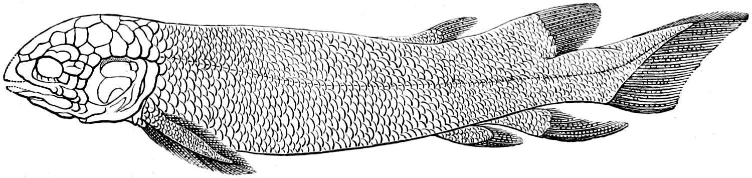
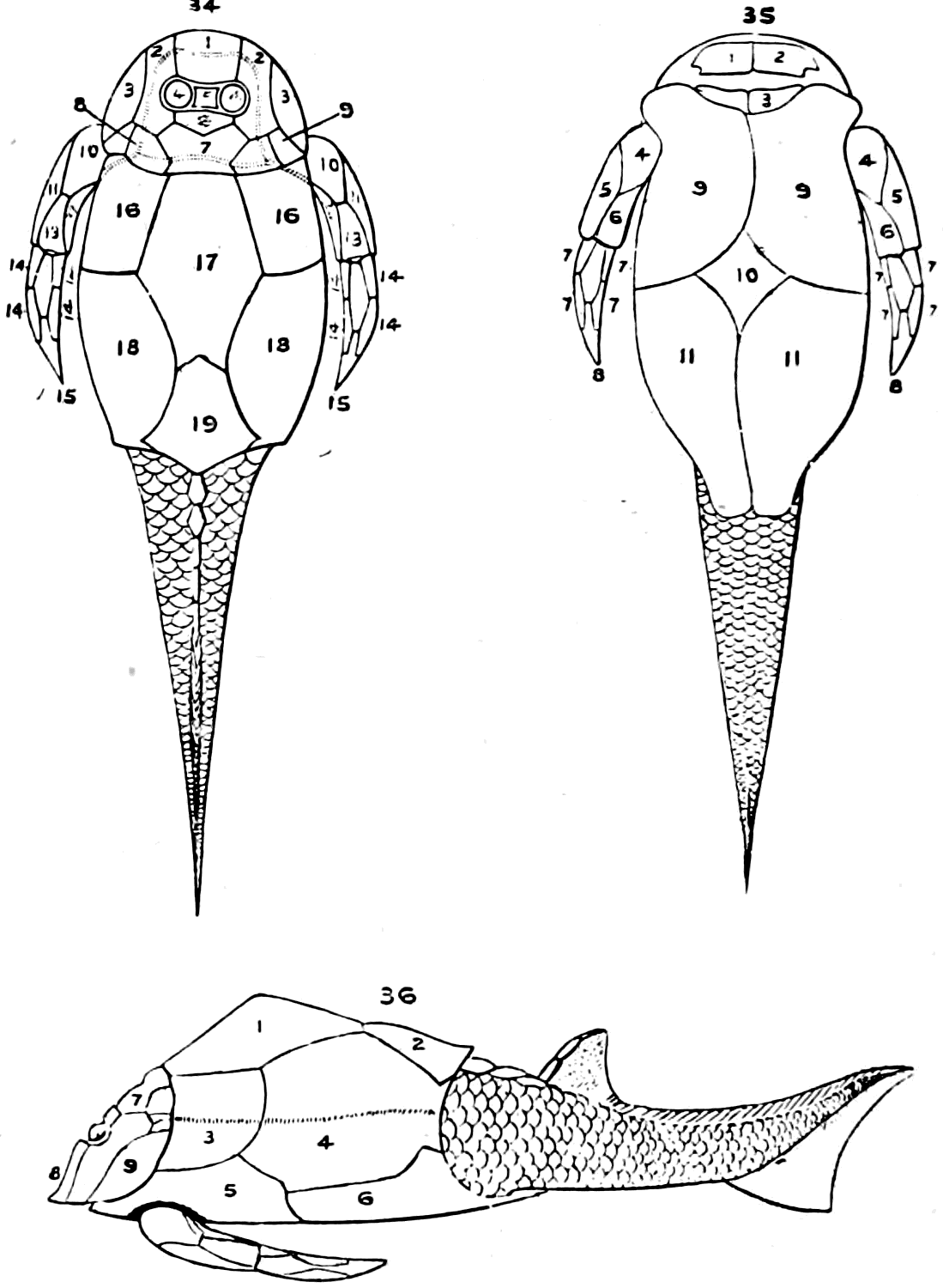
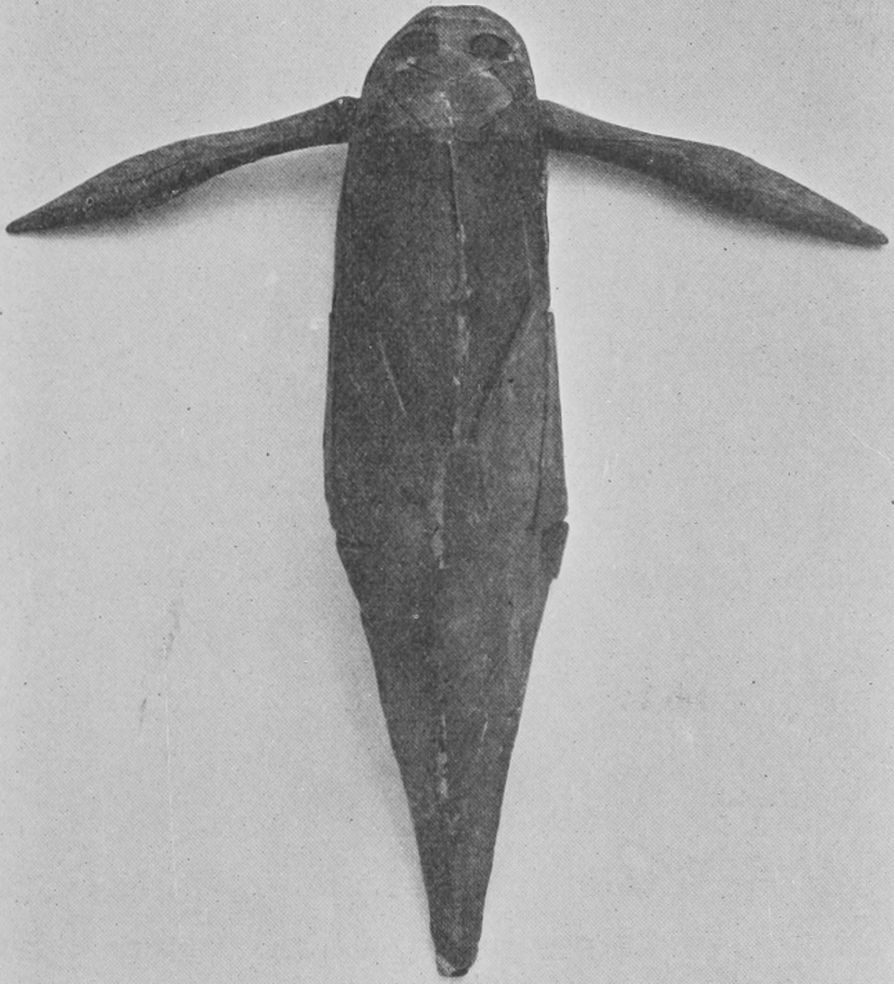
In the same rocks with Pterichthys occurs another very curious fish, the Coccosteus. This and Pterichthys were of small size only, about a foot long, but in Ohio in the United States the lower jaws and skulls of huge fishes allied to Coccosteus have been found, which must have been ten or twelve feet in length. The lower jaw of one of these (called Dinichthys), together with a restored outline of Coccosteus is shown in Fig. 185.
Very strange and curious fishes (only a few inches long) are found in still older strata—in the oldest Devonian and the Upper Silurian. One of these is called the buckler-head or Cephalaspis (Fig. 186). Its head is of the shape of a saddler's knife and the two eyes are placed near the centre. Another fish is known almost solely by the shields which covered the head or head and body, one above and the other below. This is the Pteraspis (Fig. 187). The head or head-and-body shields of these fishes and those of Cephalaspis are found in immense numbers in the hard gritty "cornstones" of Worcestershire and Herefordshire, also in Scotland. The stone is quarried for road mending, and great quantities of specimens have been found, though no other fossils occur with these fish-heads. It used to be imagined that this rock was the deposit of a great fresh-water lake, but that is not likely, since Pteraspis heads are found with marine shells in the rocks of Galicia. The curious thing is that although occasionally a tail or body of Cephalaspis covered with scales and provided with fins has been found attached to a head-shield, as in Fig. 186, yet the body or tail of Pteraspis remains unknown. The only specimen showing any trace of the hinder region of Pteraspis is one which I obtained when I was a boy (in 1864) at a quarry in Herefordshire, the workmen from whom I got it saying it was a fossilized fir-cone. As a little concession to my vanity, I have had this solitary specimen, which I gave long ago to the British Museum, photographed of the natural size (Fig. 188). It is not much to look at, but it is one of the most interesting specimens I have myself had the pleasure of unearthing. The strange thing is that it is and remains unique.
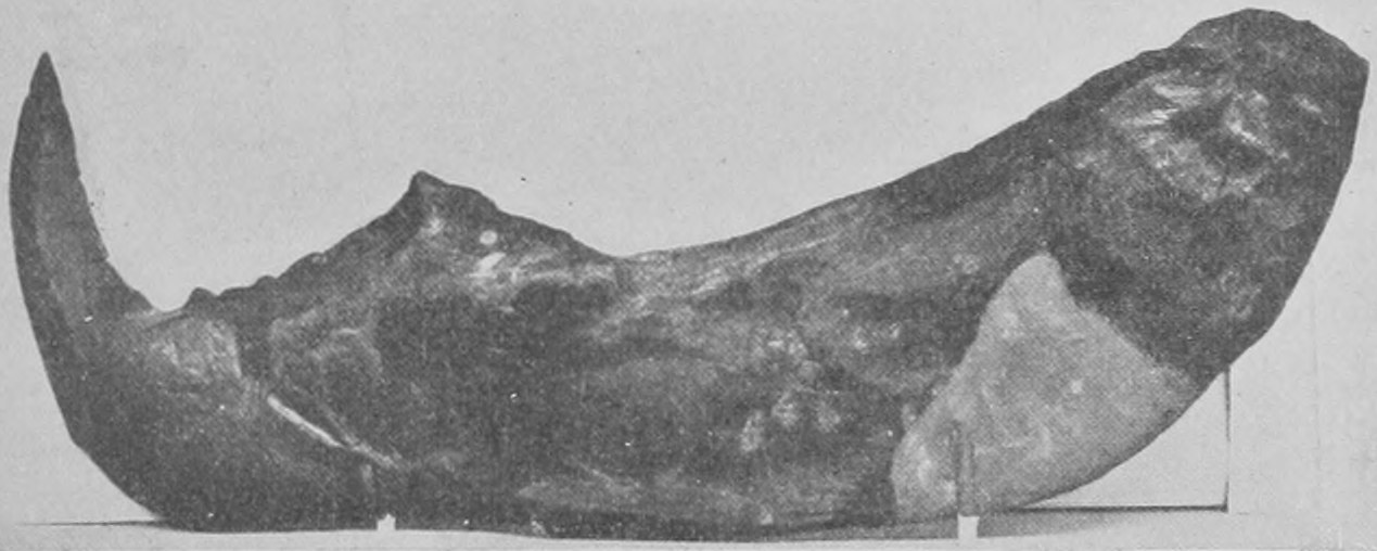
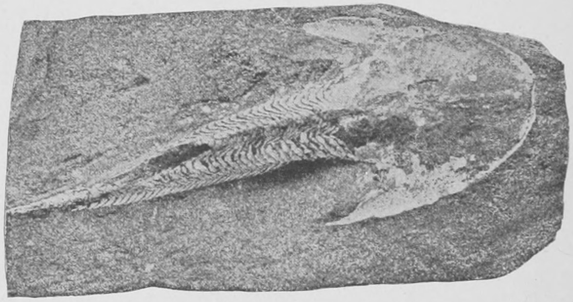
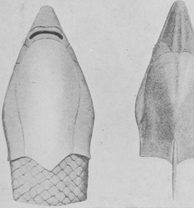
Fig. 189 is a photograph of the upper and under side of a model of the Drepanaspis, a most strange fossil fish of the same early age, allied to Pteraspis. It is prepared from the drawings of Professor Traquair, who has described the fish. Specimens of it in a crushed state preserved in the slate-rock of North Germany are in the Natural History Museum.
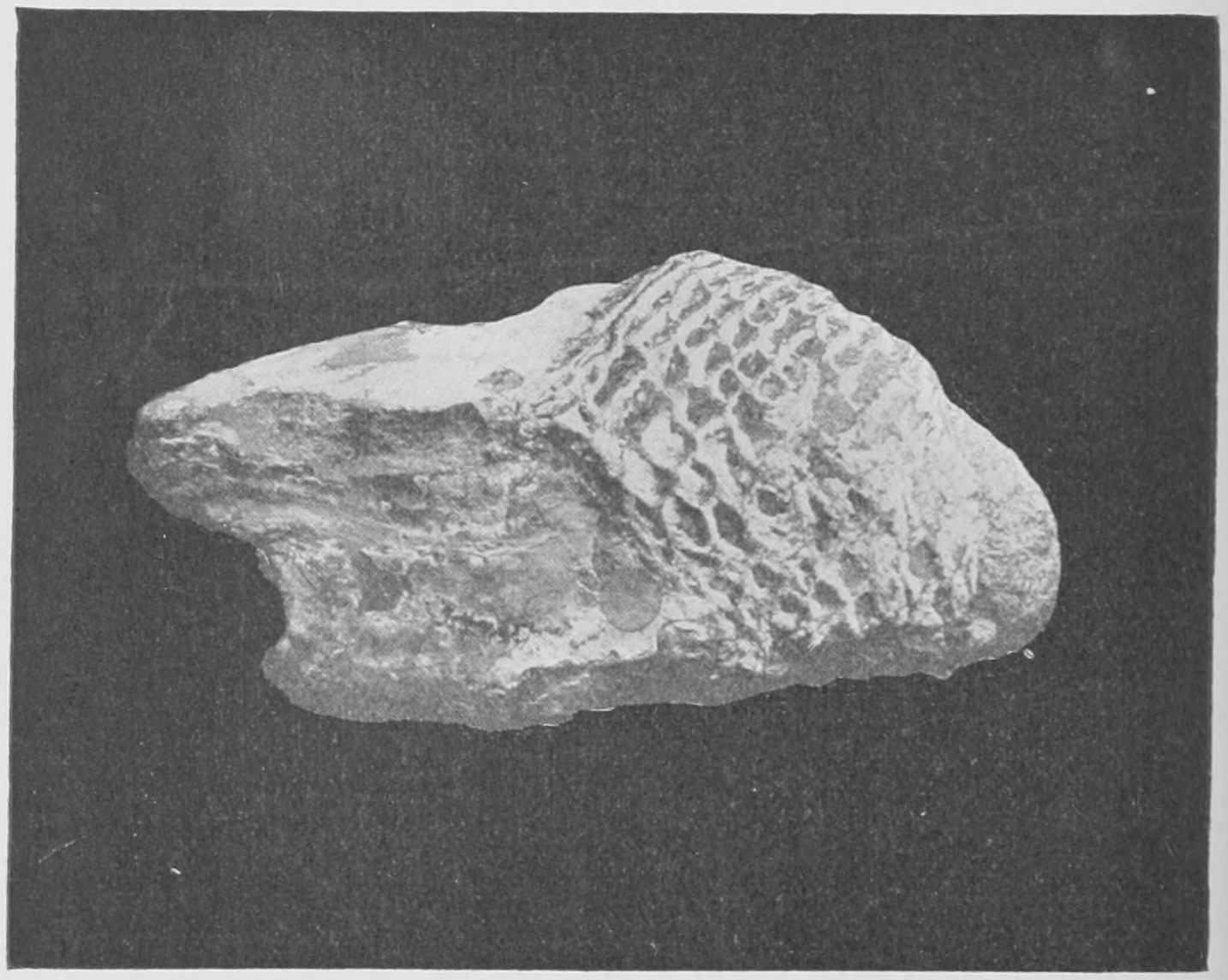
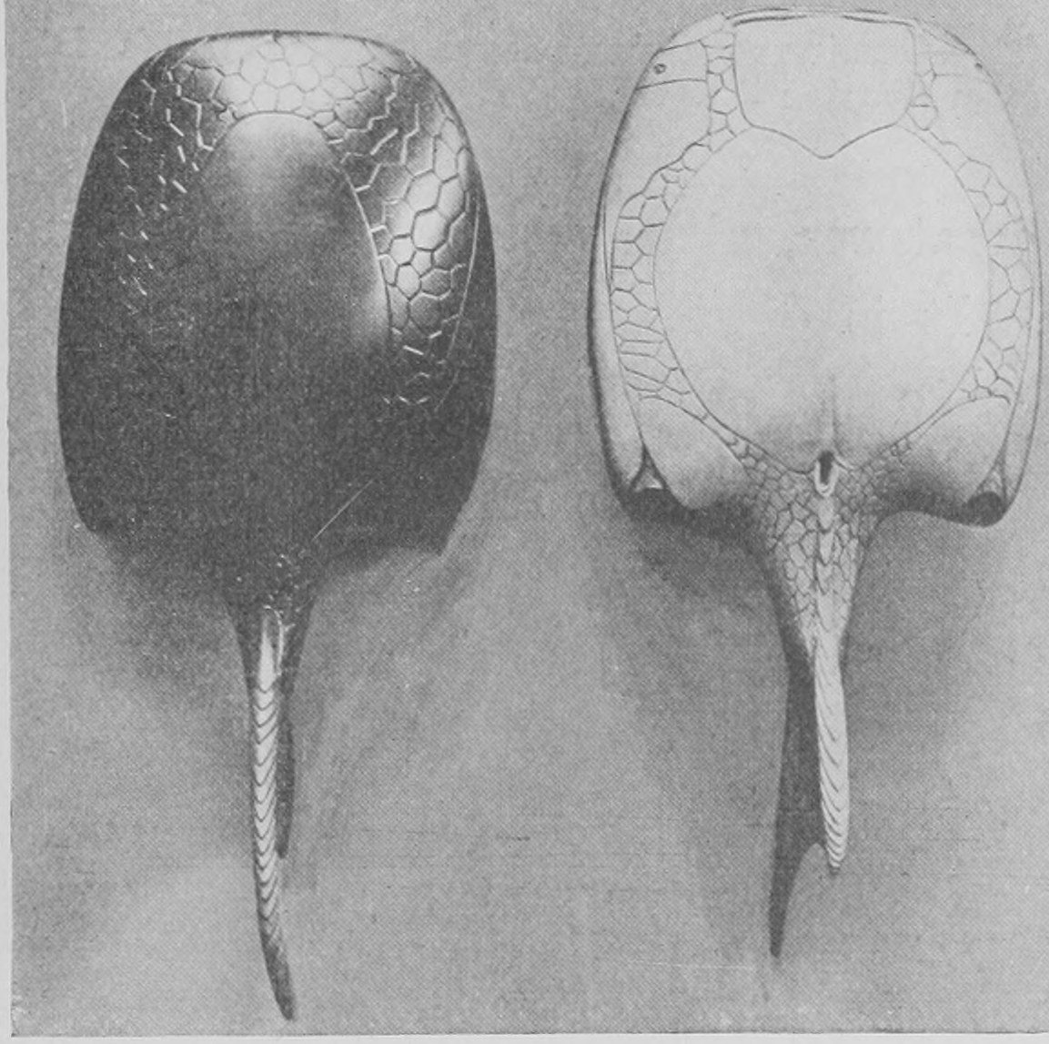
Fishes resembling this in shape have recently been found in the Silurian strata of Lanarkshire, and they, together with the curious little fishes drawn in Figs. 190, 191, are the oldest remains of fishes which have been discovered. These last two—Birkenia and Lasanius (Figs. 190, 191)—are very puzzling little creatures, with spines set in a row along the belly. It is difficult to make out back from belly or to distinguish eyes or mouth, yet they show characteristic fish tails and a scaly covering of the body. These are among the most recent discoveries and come from the Upper Silurian strata of Scotland. Specimens of these are in the Natural History Museum, but the finest series are in the Edinburgh Museum, where Professor Traquair has made a special study of the most ancient fish remains, the most ancient vertebrate remains, yet disinterred from the crust of the earth.
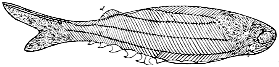
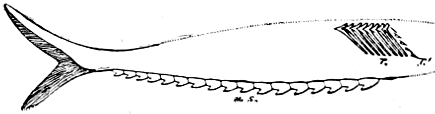
Ancient, inconceivably ancient, as are these Upper Silurian rocks, there are yet immense thicknesses below them of stratified rock, containing fossils in which no fish remains have been discovered. We must not conclude that the very curious-looking fishes of the Upper Silurian are really the actual forefathers of all later fish and of all vertebrate life. They just happen to be preserved and dug up, but probably soft-bodied fishes existed then and before that time which had no bones inside and no hard scales outside, and so have left no sign, in the rocks, of their existence. The Upper Silurian strata are, as you will see by looking at the Table of Strata on p. 60, just halfway down in the thicknesses of rocks, between the present river gravels above and the Cambrian beds with the oldest known fossils (certain Trilobites) below.
We will revert to the Trilobites directly; but before leaving the extinct fishes I wish to mention the great fossil sharks of the late Tertiaries (Miocene and Pliocene). These we know by their teeth; enormous shark's teeth are found which are three times the length of the teeth of the biggest living sharks on record, as shown in Fig. 192. These teeth are found in beautiful preservation in Malta, in the Antwerp sands, in Maryland, U.S.A., and in Suffolk in England. In Suffolk they occur in the same wonderful bone-bed of the Red and Coralline Crag (see Fig. 192A), from which we get the teeth of mastodon, rhinoceros and tapir. It seems to be a correct conclusion that this huge shark (Carcharodon megalodon) was nearly one hundred feet in length, since its teeth were fully three times the length of an almost identical recent shark (Carcharias rondeletii), which measures thirty feet in length.
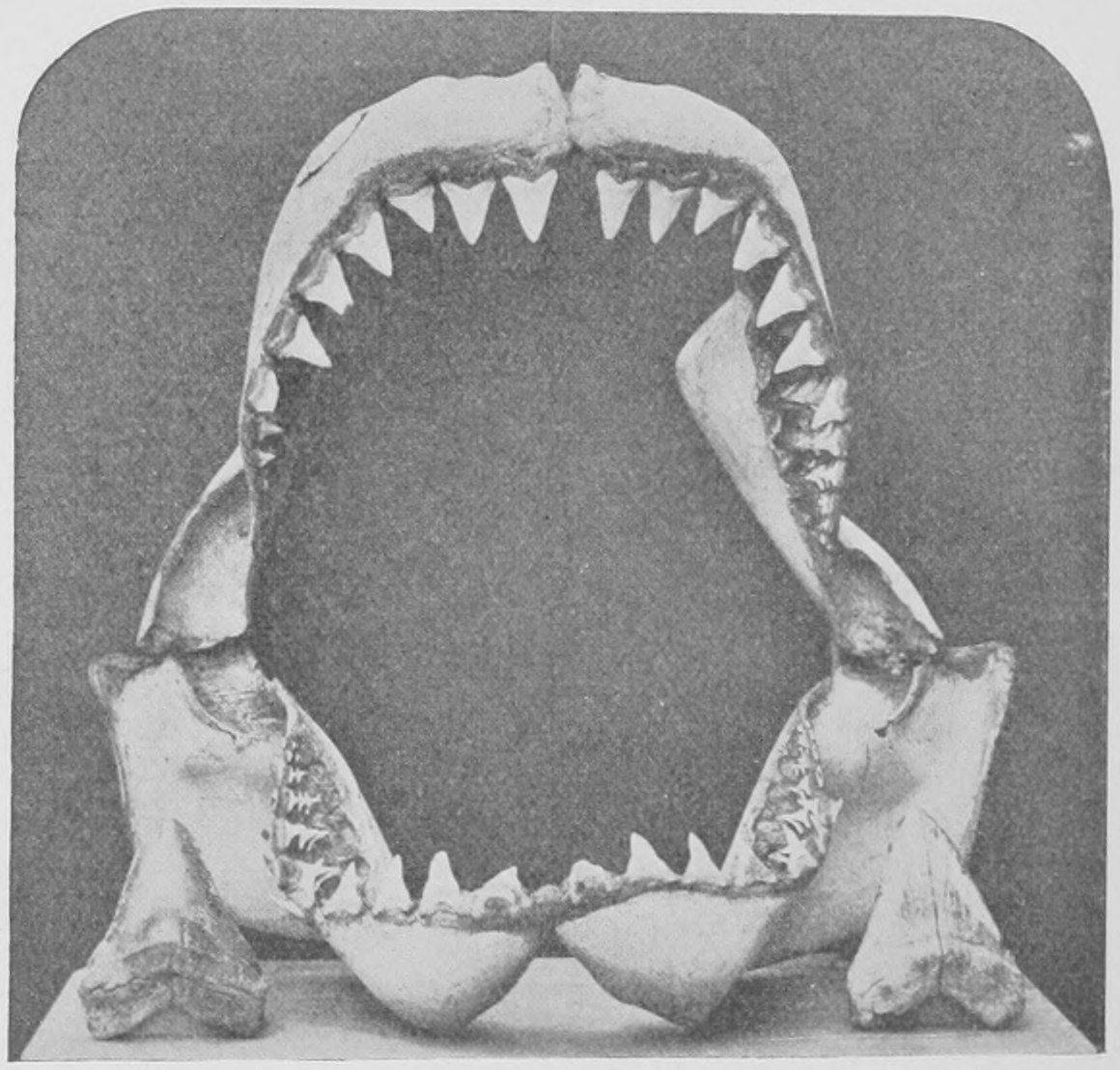
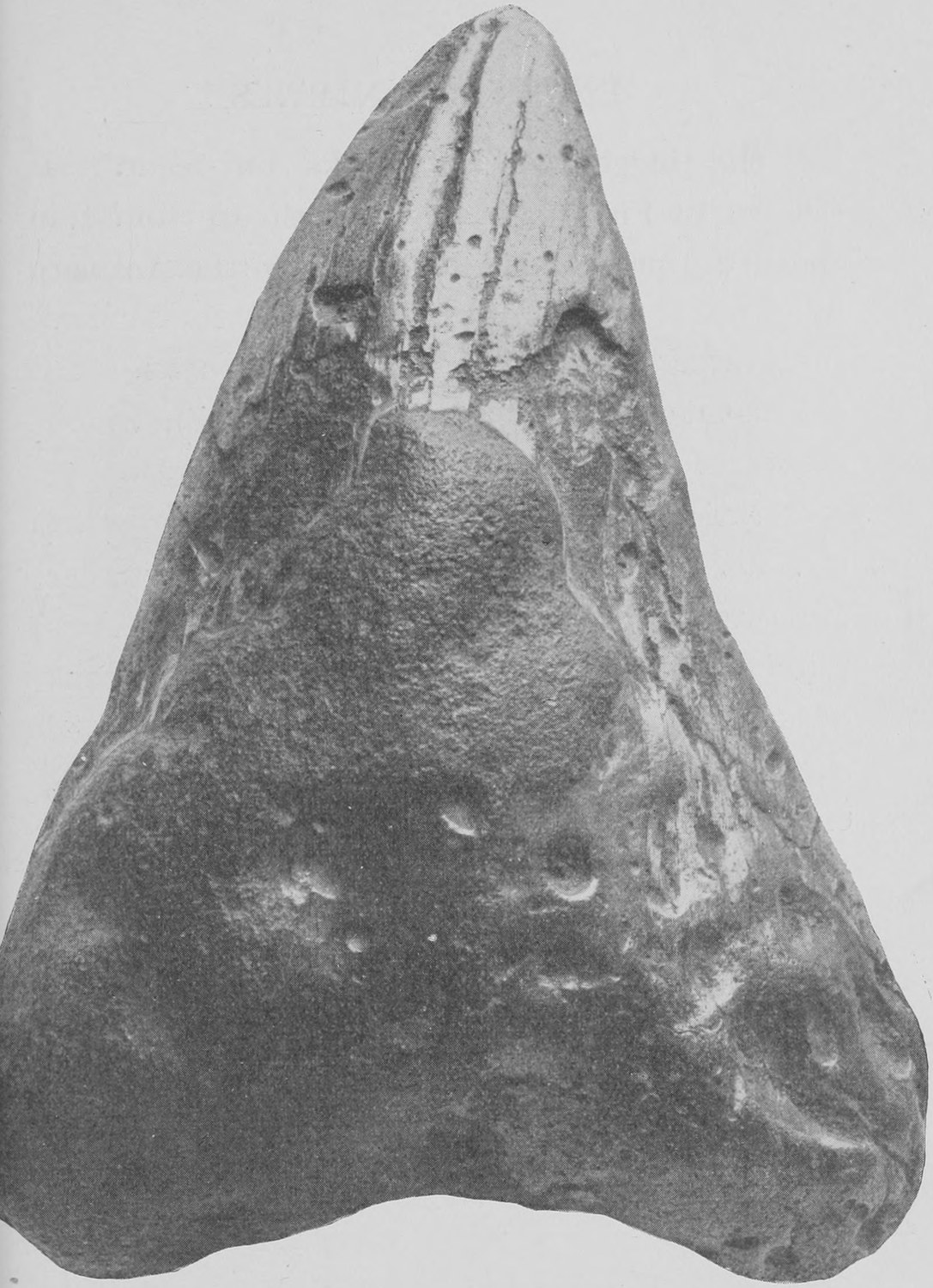
"Extinct animals" include, as must be obvious at once, a vast number of smaller creatures besides the vertebrate Fishes, Amphibians, Reptiles, Birds and Mammals. Rocks occur containing thousands, even millions, of shells of Molluscs (whelks, bivalves, etc.) crowded together in a space of a few feet. Remains of minute shrimps are equally abundant, and whole mountains are built up of rock formed by the coral or calcareous skeleton of minute polyps resembling our sea-anemone. Many of these are very peculiar forms, unlike those now living. Others, again, are remarkable for the fact that though found in the most ancient rocks they yet closely resemble creatures still living to-day.
We will now glance at a few of the more remarkable "fossils" of these lower or simpler kinds. (See the table of classes on p. 56.)
In the Jurassic strata and in the Greensand and Chalk wonderful coiled shells are very commonly found which have been compared by the country-folk to petrified snakes and to the coiled horns of the ram. These are the so-called Ammonites (Fig. 193), of which there are a great number of different kinds, some as big as five feet in diameter. When cut across they are seen to be divided into a number of chambers internally. In fact, their structure is the same as that of the beautiful shells of the Pearly Nautilus (Fig. 193A), which to-day lives in the Indian and Pacific oceans. The chambers in the shell of the pearly nautilus contain gas and act as a float, whilst the animal lives in the last chamber (Fig. 194). There are only some three or four species of pearly nautilus now living, and they represent a vast variety of extinct creatures which comprise not only the Ammonites but the more ancient Goniatites. Some of these extinct allies of nautilus, such as the Orthoceras, were not coiled but quite straight; others were loosely coiled, as is the Ancyloceras shown in Fig. 195, and others were twisted into elongated spires (Turrilites).
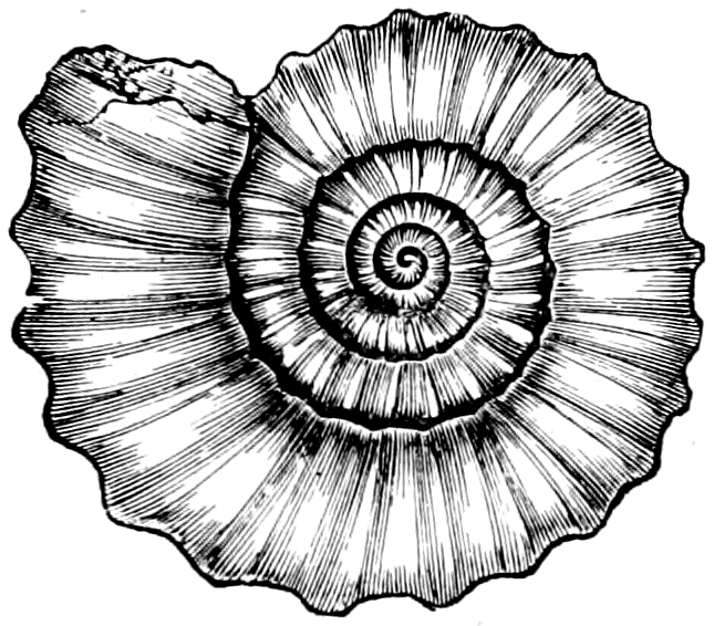
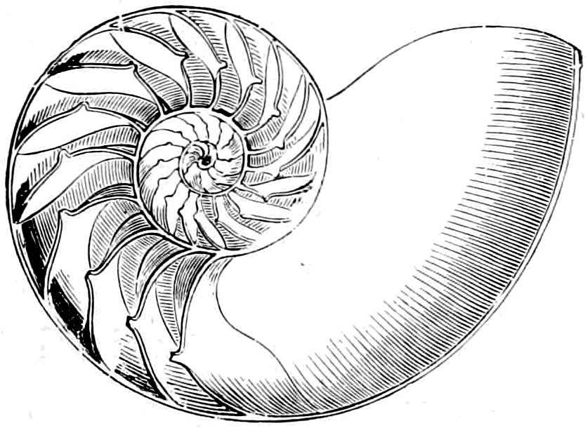
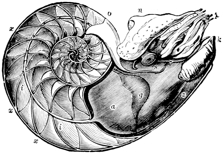
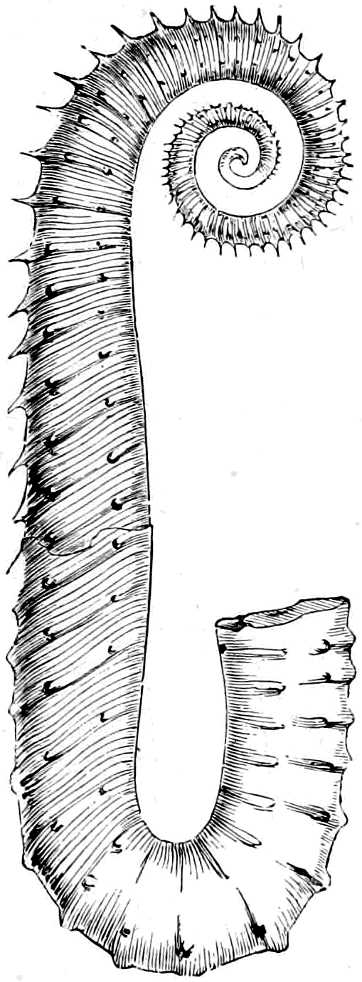
The creature which lived in these shells was similar to a cuttle-fish (as we know from examination of the animal of Nautilus), and belonged to the class Cephalopoda of the great group Mollusca. The Molluscs include, besides these, the whelks, snails, mussels, clams and oysters.
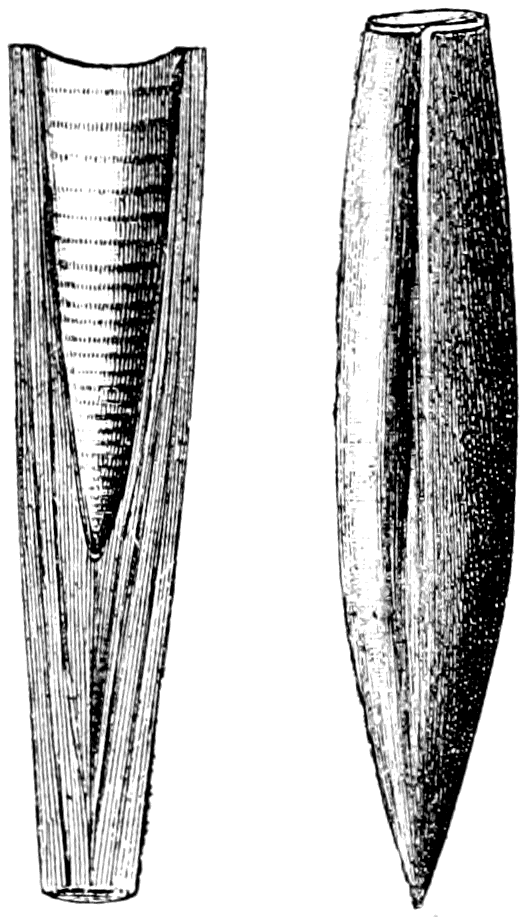
A celebrated fossil which is the internal shell or "pen" of a kind of cuttle-fish is that known by the name "Belemnite" (Fig. 196). These fossils are called "thunder-bolts" in some parts of England, where they are sufficiently common in the clay and shale to attract attention. They are found only in the Jurassic and Cretaceous formations. In fine clay specimens occur showing the soft parts of the sort of cuttle-fish in which they were formed (see Fig. 197). They are of the same character as the "cuttle-bone" of the living cuttle-fish and the pen of the squid (Fig. 198), but are more solid and heavy.
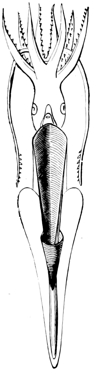
The oldest fossils which are known are found in the Lower Cambrian rocks (see Table of Strata, p. 60), and are the remains of small marine creatures, which were, however, by no means very simple in structure. One of these is the Lingula davisii (Fig. 199), from the Lingula flags of Wales. Only the simple oval shells are known, but they are almost exactly like the shells of a marine animal which is still found living in immense numbers on the shores of the warmer oceans. The living owners of these shells occur in great numbers burrowing in sand and have a very highly complex structure and red-coloured blood. It is indeed a most remarkable fact that the remote fossil shells of the lower Cambrian strata should be identical with those of a living animal of a high rank in the scale of structure. Not only is that the case, but in all the deposits above the Cambrian we find the shells of Lingula, so that we must conclude that Lingula has been existing in the seas of this earth, with very little change in form, ever since the Lower Cambrian times.
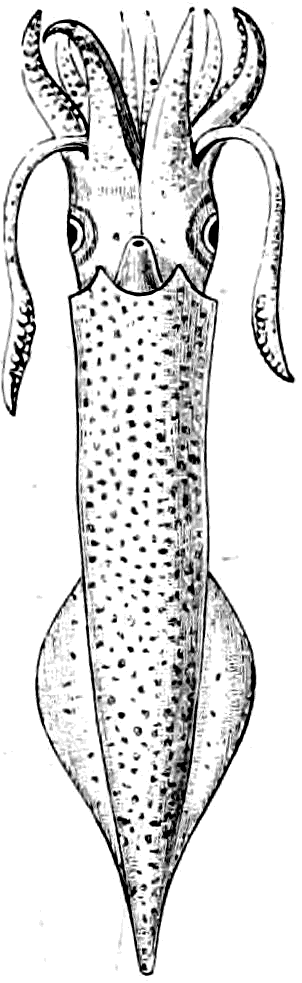
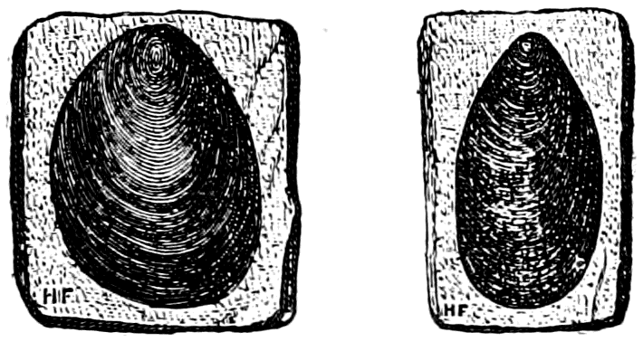
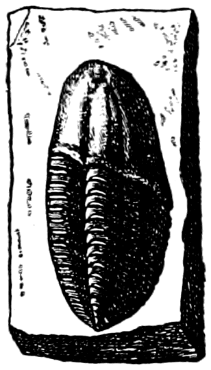
Another class of fossils which are equally ancient are the Trilobites (Fig. 200). These are well-marked forms with ringed or jointed bodies divided very often into three longitudinal lobes; hence the old name Trilobites. An immense number of different kinds of Trilobites are known and classified, but they ceased to exist in the Permian period (see Table of Strata, p. 60). For a long time the legs of these creatures were unknown; they have only been found within the last ten years. Mr. Beecher, of the United States, discovered them in one particular kind—the Triarthrus becki (Fig. 201). Some people consider these animals to be allied to the wood-lice or other crustacean shrimp-like forms now living. But it seems most probable that they were a primitive marine group allied to the scorpions, spiders and king-crabs (the Arachnida).
It is a fact of very great significance that the earliest fossils yet discovered are the remains of very highly developed animals, by no means near the beginning of animal life. It is indeed a reasonable supposition that the earliest forms of animal life must have preceded the Cambrian Trilobites and Lingula by as long a period as these latter precede the animals living to-day. Apparently the soft-bodied animals which preceded the Cambrian fossils have not left any remains in the rocks below the Cambrian or their remains have been destroyed by chemical and structural change in those most ancient deposits.
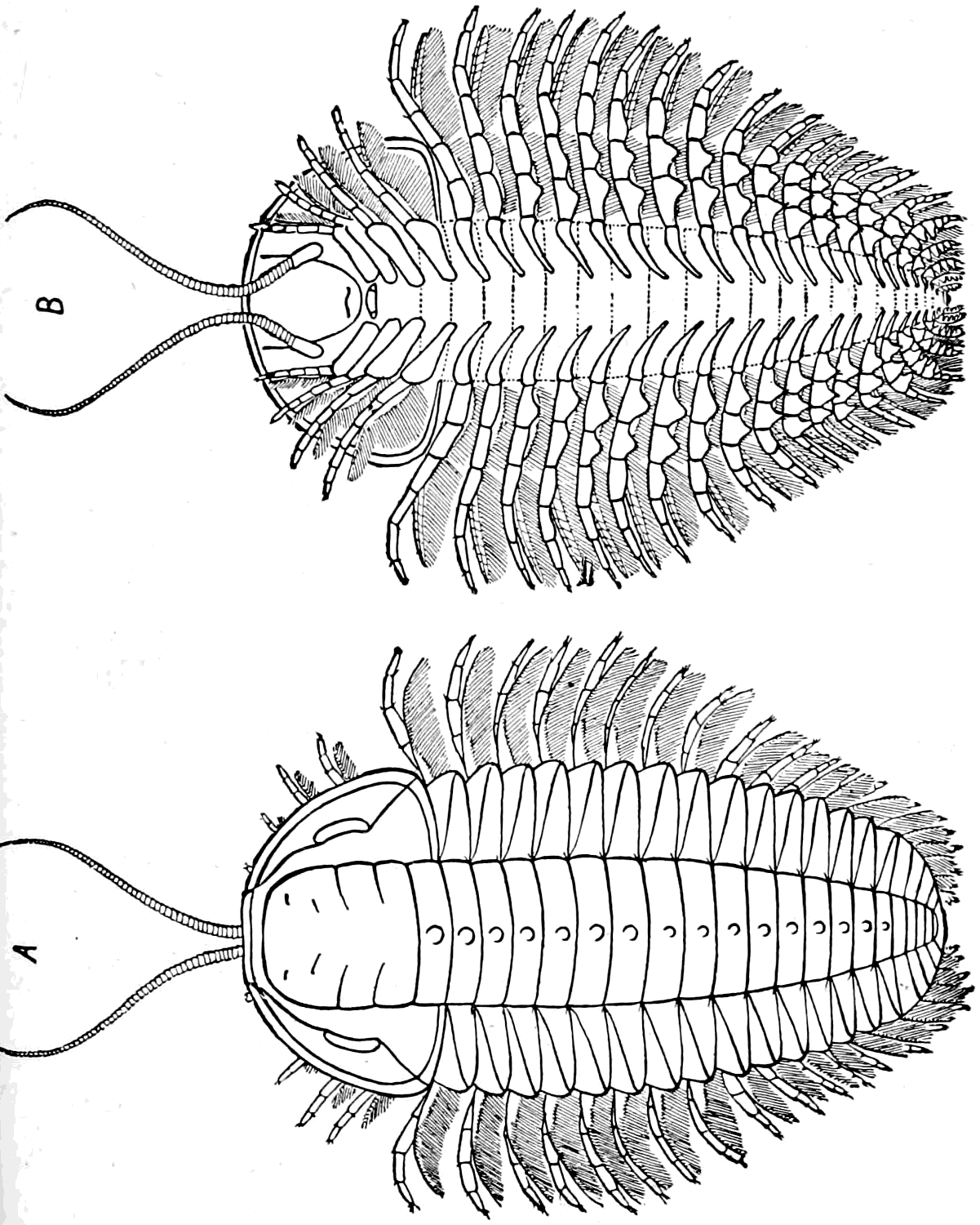
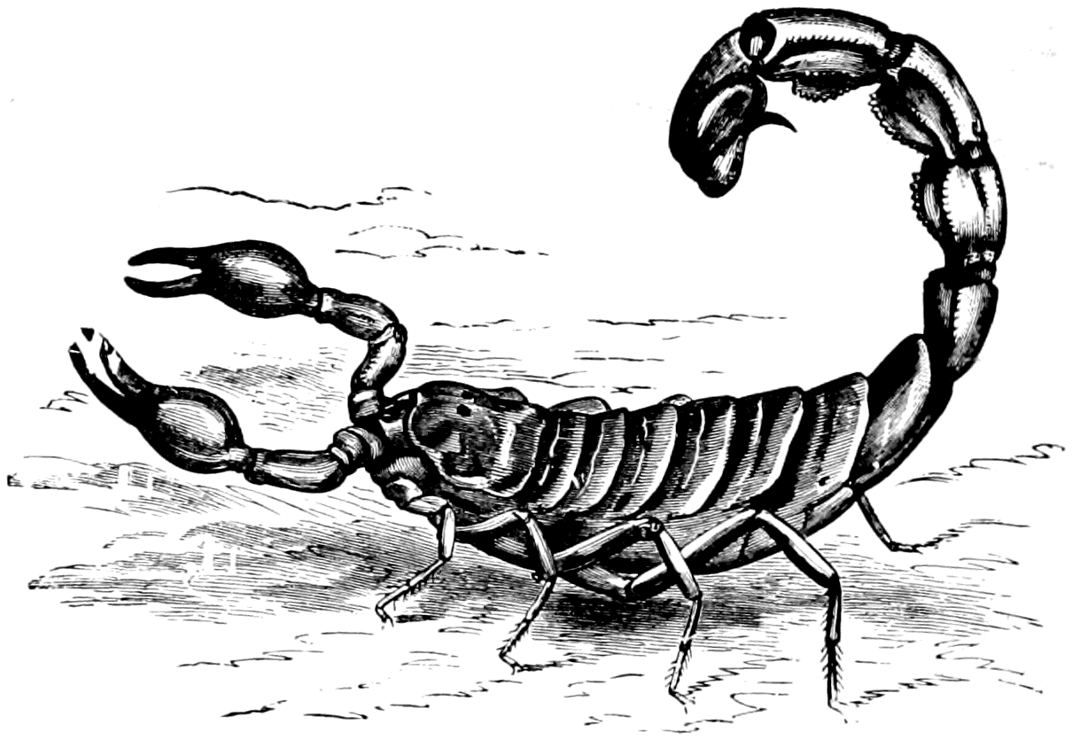
The Scorpion itself (Fig. 202) is a very ancient and important animal which so far impressed the imagination of even the earliest civilized men, that they named one of the constellations after it. Some hundreds of distinct species of scorpions are known as living at the present day in various parts of the world. In the Carboniferous strata we find fossil scorpions hardly differing at all from those now alive, and even in the Upper Silurian we find a scorpion (Fig. 203), which would be recognized at once by a child as being a true scorpion. It, however, seems probable that whilst modern scorpions are terrestrial, and breathe air by means of lung-sacs, the Silurian scorpion was aquatic. This is indicated by its thick crab-like legs with strong pointed end-joints (Figs. 204 and 205).
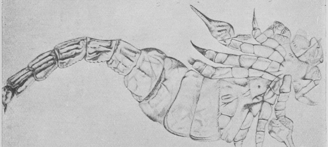
Besides the Silurian scorpion of undoubted affinity to modern scorpions, we find in the Silurian and Devonian rocks remains of enormous aquatic scorpion-like creatures, sometimes four or five feet in length (Figs. 207, 208, 209). These are known as the Eurypterids (Pterygotus, Stylonurus, Eurypterus, etc.). They had six legs like the scorpion, of which the anterior carried nippers in some instances. The great spine at the end of the body is the representative of the scorpion's sting, whilst they agree with scorpions in the position and character of the eyes and in the number of segments or rings which build up the body and the head.
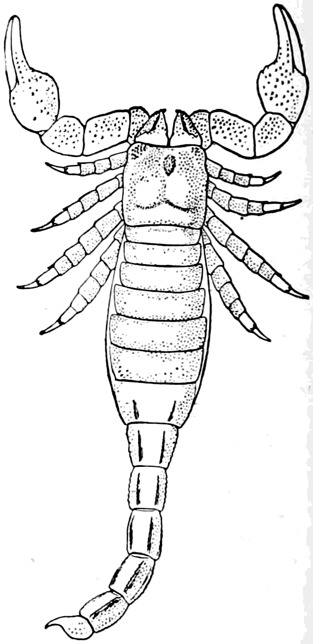
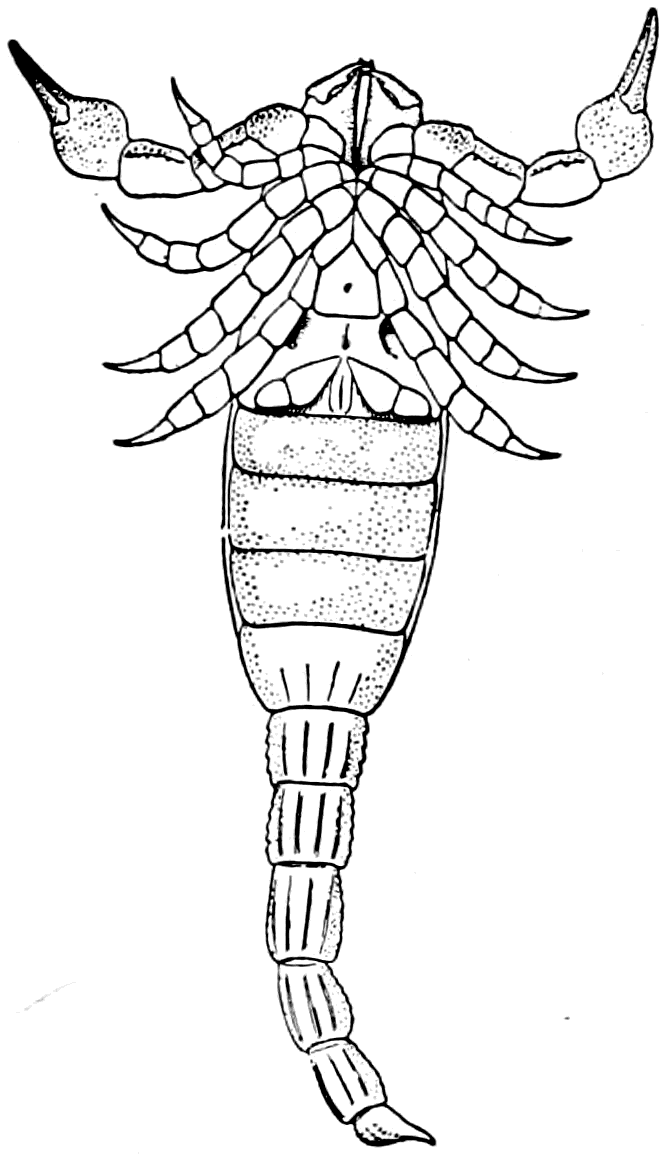
A very interesting animal which is still alive (but is also found in ancient rocks) connects the scorpions with the great extinct Eurypterids and also with the Trilobites. This is the King-crab (Figs. 210, 211, 212), which is not a true crab—that is to say, a member of the class Crustacea—but is a sort of marine scorpion with shortened tail (though having a long sting-like spine at the end of its body)—a member of the class Arachnida. Its legs, six in number (Fig. 212), are singularly like those of the scorpion, and in a great number of minute details it agrees with scorpions (see Fig. 213) and differs from crabs. It is the only surviving representative of the aquatic ancestors from which the modern air-breathing scorpions and spiders have been developed.
From amongst all the great variety of extinct invertebrate animals, I select for our last illustrations and descriptions a few of the beautiful stone-lilies or Pentacrini, or Encrinites as they were long ago called. They have a very interesting history, for they were known as fossils as long ago as the seventeenth century, many years before they were found in the living state. They are a kind of star-fish, with long delicate arms attached to a central cup or body which is mounted on a jointed stem, which is often of great length. Several kinds are shown in the figures 214, 215, 216. The fossil remains which we find are the hard internal skeleton, consisting of carbonate of lime, which was a very prominent feature in their structure. It is of the same nature as the hard box-like skeleton of the sea-urchins, which, with the star-fishes, form the great group called by naturalists the Echinoderma.
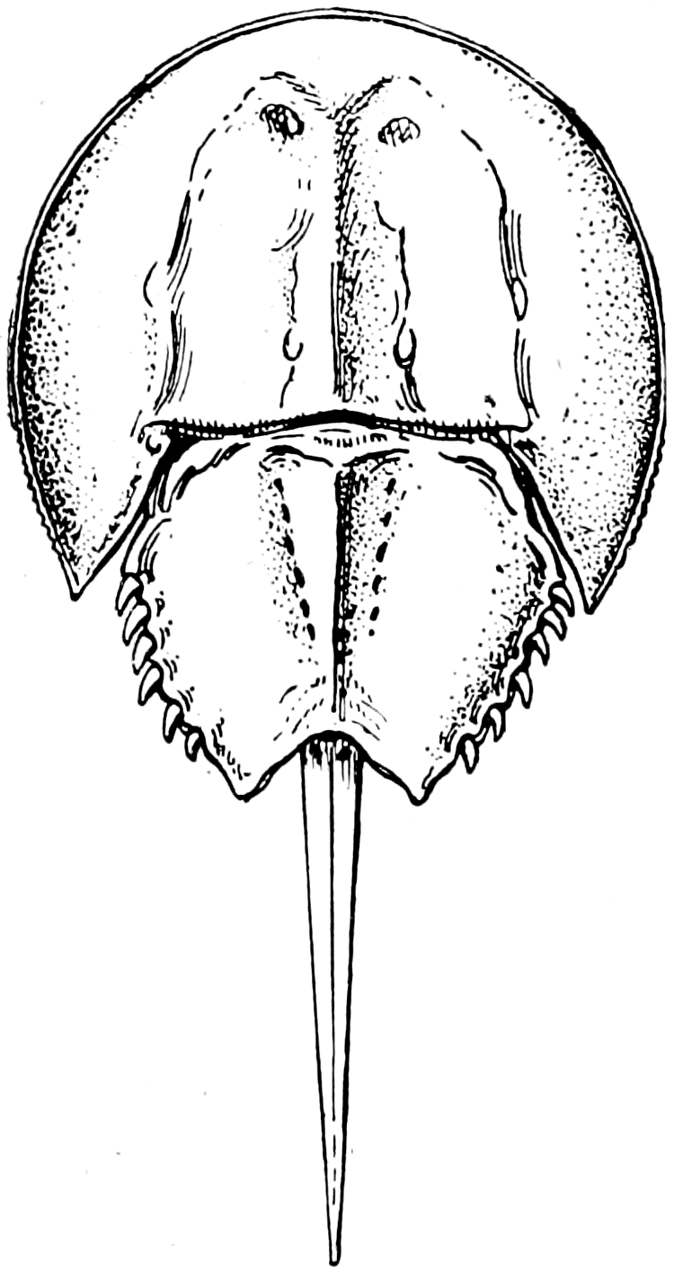
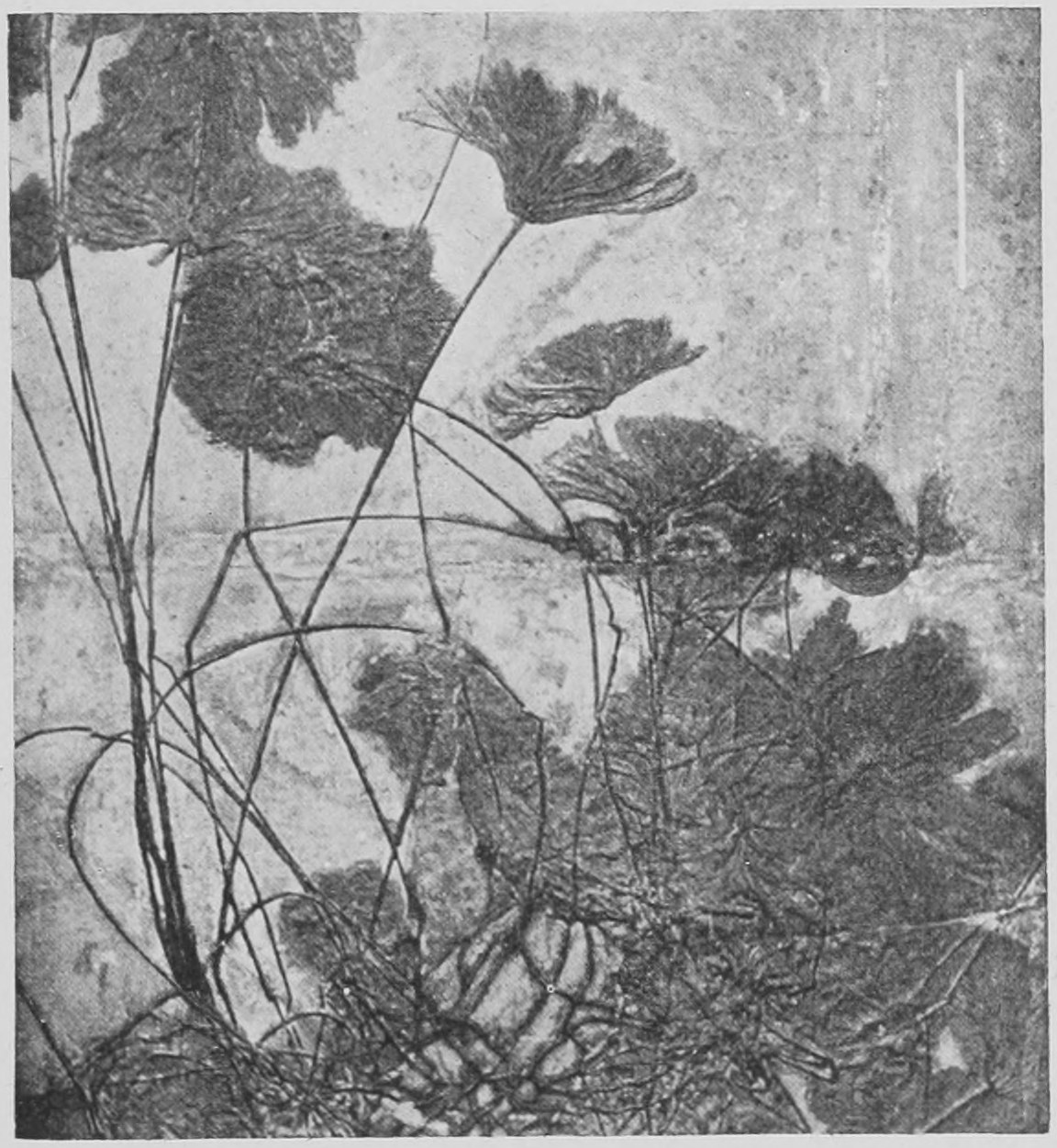
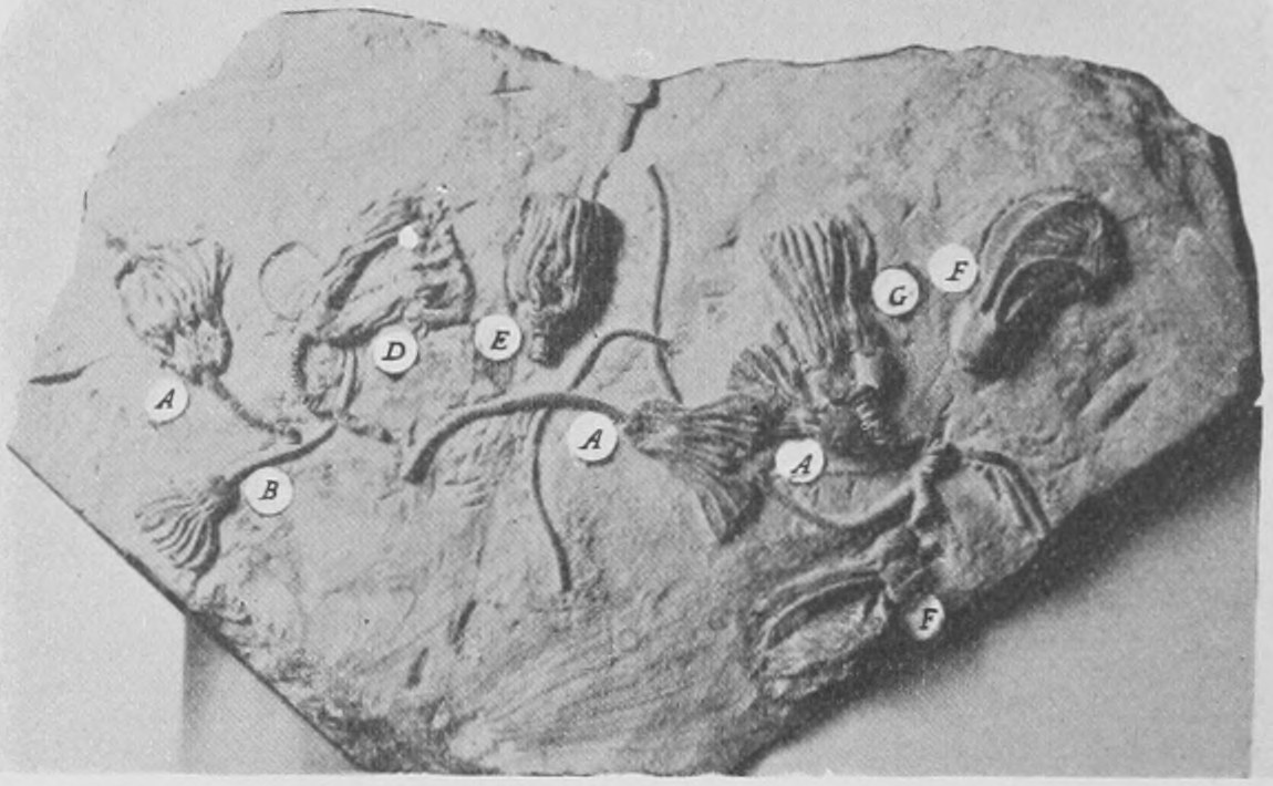
In the eighteenth century a specimen of a living Encrinite or stalked star-fish was discovered in the deep water off Martinique in the West Indies, and was brought in a dried condition to Europe and described as a "sea-palm-tree." For a long time such specimens were very rare and difficult to obtain, but now a great number have been dredged up in deep water in different parts of the world. Still there are only a dozen or so of different kinds or species of the Encrinites still living, whereas in all the older rocks we find their remains often in great profusion. Many hundreds of extinct kinds are known and they occur as far back as the Cambrian rocks and are wonderfully varied and abundant in the Silurian, Devonian and Carboniferous (Fig. 215). Some of the finest are found in the Jurassic strata (Figs. 214 and 216).
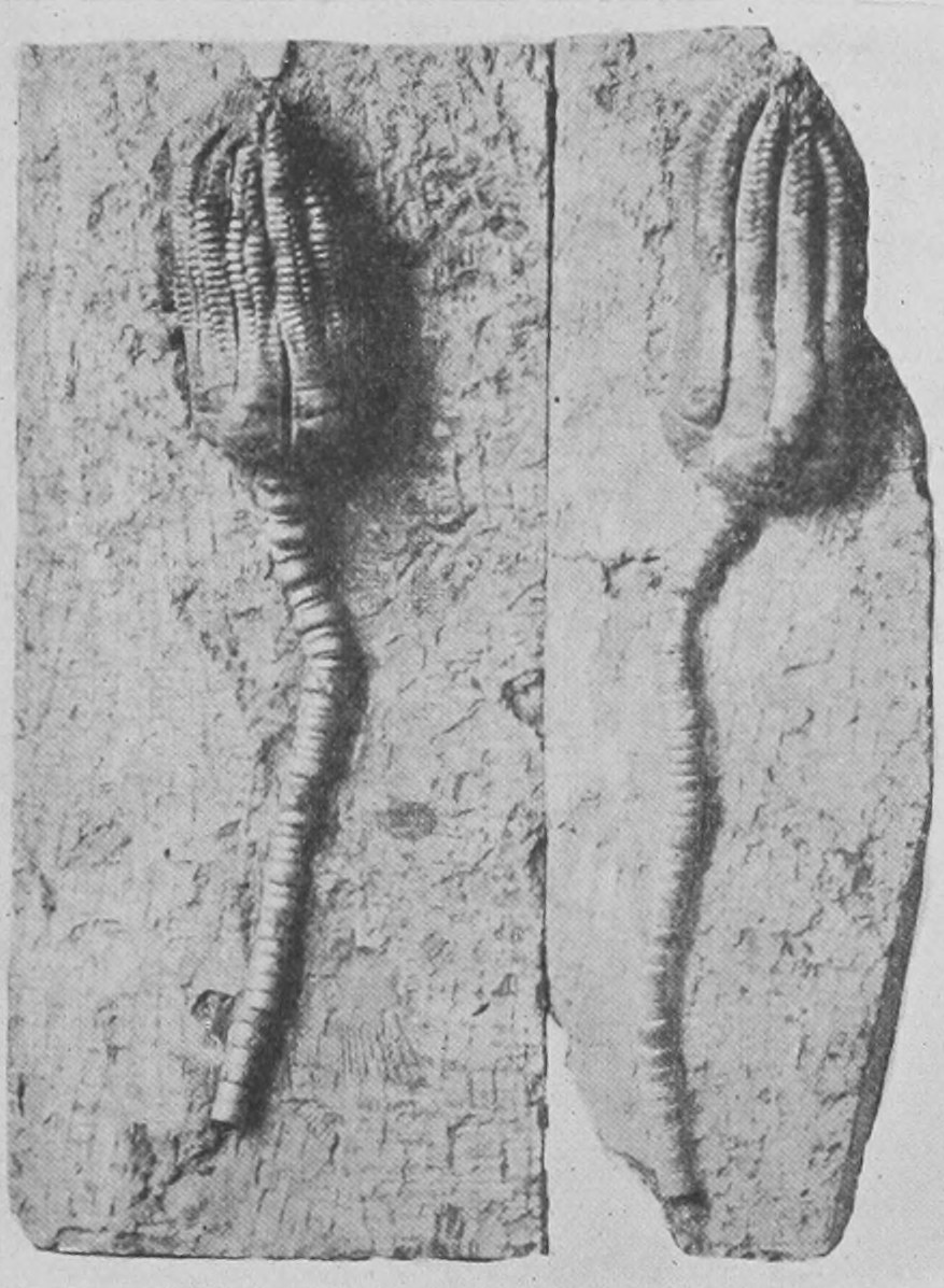
A very interesting discovery in regard to the Encrinites was made by a celebrated English naturalist, Vaughan Thompson, in 1836, who was an army surgeon and quartered at Cork, where he studied the marine animals of Queenstown harbour. He found out many new and important things by watching the growth from the egg by means of the microscope of barnacles, star-fishes and sea-moss, which he kept alive in small glass vessels.
Vaughan Thompson first of all discovered in the sea at Queenstown a minute Encrinite, not a third of an inch long (Fig. 217), and to this he gave the name Pentacrinus europœus. The large one from the West Indies was at that time the only other living Encrinite known, and was called Pentacrinus asteria.
This was a sufficiently astonishing discovery, but more was to come. Vaughan Thompson found in the next place that the body of his little Pentacrinus europœus grows larger and larger whilst the stalk shrivels and ceases to grow, so that the animal becomes detached and swims away freely by the movement of its arms. It grows up to be a well-known star-fish, the feather-star or comatula (Fig. 218). At first this history was not believed, and the Royal Society of London refused to publish Vaughan Thompson's account of what he had seen. But it was soon fully established. The little Pentacrini were bred in glass jars by many observers from the eggs of the feather-star, and all of them were seen to proceed after a certain time to produce freely-swimming little star-fish. Thus it was proved, what indeed is clear enough from the structure of its feathered arms and other parts, that the feather-star or comatula is unlike other star-fishes and is a Pentacrinus (or stone-lily or Encrinite or Crinoid) which has lost its stalk. And all the time that the naturalists of 250 years ago were disputing as to the real nature of the stone-lilies found in the rocks, little stone-lilies a quarter of an inch long were being abundantly produced every year close to hand in the sea on the rocky shores of England and France and in the Mediterranean. Whilst the first recent unfossilized Pentacrinus seen by naturalists was brought all the way from Martinique, any number of a minute size were to be found living on our own shores. But these European Pentacrini escaped observation on account of their minute size and the sudden dwindling and loss of the stalk. Only in its very young stage does the common feather-star of to-day retain the most remarkable characteristic of its remote Cambrian ancestors, the stalk. But it does for a brief week or so, whilst almost invisible to the unaided eye, possess a well-grown stalk by which it is fixed just as were its remote forefathers by their splendid waving stems many feet in length.
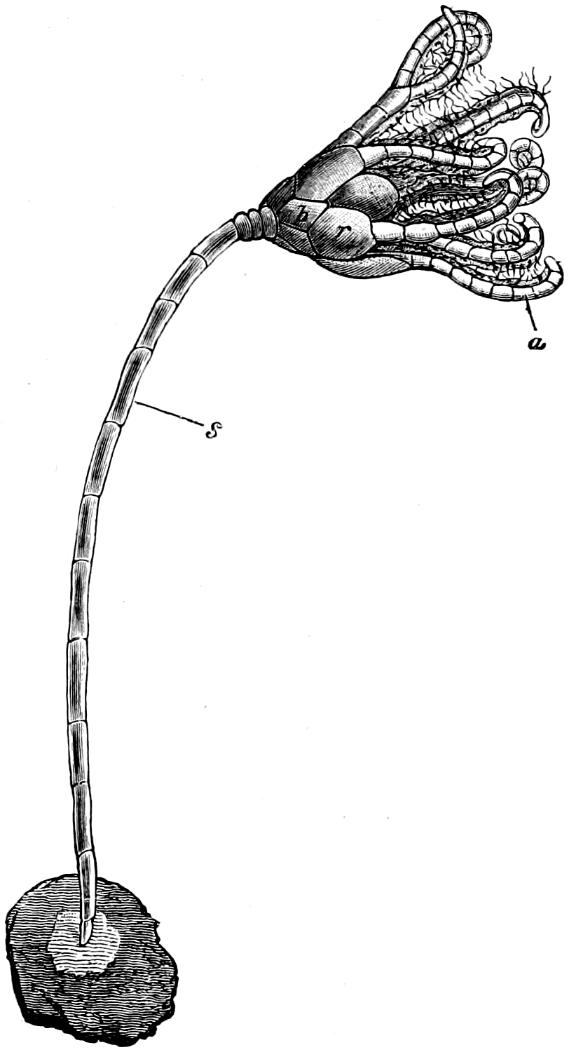
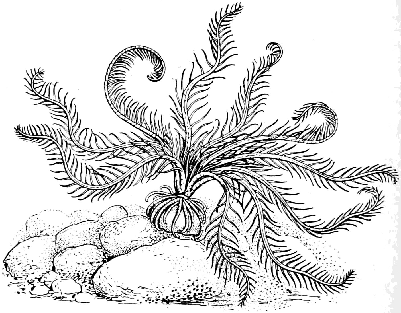
In these pages I have only been able to bring to the reader's notice a few of the marvellous and delightful things which we know as "fossils"—only a very small selection of what is known about extinct animals. I have avoided going into much detail and using more technical terms and long names than is absolutely necessary. It is impossible to speak of these things without mentioning their names; and though it is true that a fossil "called by any other name" would still be full of interest, yet we must have a definite name by which to speak of each kind of animal and each kind of rock. If one's interest is aroused in these astoundingly ancient and curious remains of extinct creatures, it becomes after all no very difficult matter to remember their names and to distinguish them from each other as well as to recognize them when we come across the names in books or museums. To learn more than the few facts which I have so briefly stated in these pages, the reader should visit many times the Natural History Museum, see the actual specimens, and by the aid of the illustrated guide-books get to know more details about them. And if he or she have the chance and can go and hunt in some of the quarries or cliffs which are so often full of fossils, an endless delight and a health-giving pursuit is the prospect before him or her. Fossil-hunting with hammer and chisel and a bag to be laden with specimens, is splendid exercise, and, if skilfully conducted, an exciting form of sport. Even within reach of a Londoner's day there are the brickfields of Ilford and Grays, where I used to get remains of mammoth, rhinoceros and such beasts; there are the chalk and tertiary strata of Charlton in Kent full of fossils; the Red Crag pits of Suffolk; the oolites of Oxford; the sponge-gravel of Farringdon. A very little longer journey brings the fossil-hunter to the Isle of Wight, which used to be, and I doubt not still is, a magnificent preserve of Eocene, Greensand and Wealden fossils; and not further off in length of journey are the Malvern hills, with Silurian and Devonian strata exposed in quarries and railway cuttings, teeming with fossils. And there is always the chance—a good sportsman's chance—of finding "something new" if one understands the business and is never wearied of digging in sand and clay, and hammering the rock, and hunting up quarrymen and those delightful people—rarer now than they were forty years ago—the local naturalists. I hope that many, if not all of my readers, may be incited by the accounts and pictures of extinct animals which I have given in these pages, to become "soldiers of the hammer," as Sir Roderick Murchison used to call us, and collectors of fossils—and if blessed by good fortune, discoverers of things as yet unknown to man.